2011年1月31日 星期一

CT for Acute Aortic Syndrome


CT Imaging for Acute Aortic Syndrome
Cleveland Clinic Journal of Medicine 2008; 75(1):7-24

Diagnostic strategy for acute aortic syndrome


Acute aortic syndrome

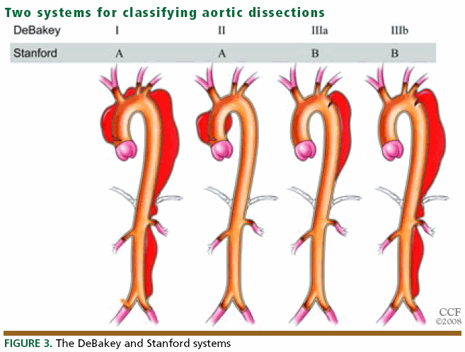
1.  Acute aortic dissection

2. Intramural hematoma
Acute intramural hematoma is easily recognized in CT without contrast by the higher Hounsfield-unit value of the blood products in the wall in comparison with the flowing blood in the lumen, eccentric aortic wall-thickening and displacement of intimal calcifications.
3. Penetrating atherosclerotic ulcer

4. Unstable thoracic aneurysm
An aortic aneurysm is defined as a permanent dilation at least 150% of normal size, or > 5 cm in thoracic aorta or > 3 cm in abdominal aorta.
CT signs of imminent rupture include a high-attenuating crescent in the wall of the aorta, discontinuous calcification in a circumferentially calcified aorta, an aorta that conforms to the neighboring vertebral body (“draped” aorta), and an eccentric nipple shape to the aorta.

2011年1月25日 星期二

The Management of Pancreatic Trauma in the Modern Era


The Management of Pancreatic Trauma in the Modern Era
Surgical Clinics of North America. Volume 87, Issue 6 (December 2007)

Diagnosis
  • Grading system
  • Serum amylase levels
  • CT
  • ERCP
  • DSS MRCP
  • Exploratory laparotomy
Pancreas Organ Injury Scale of the American Association for the Surgery of Trauma

Grade
Injury
Description
 I
Hematoma
Minor contusion without duct injury

Laceration
Superficial laceration without duct injury
 II
Hematoma
Major contusion without duct injury or tissue loss

Laceration
Major laceration without duct injury or tissue loss
 III
Laceration
Distal transection or parenchymal injury with duct injury
 IV
Laceration
Proximal transection or parenchymal injury involving ampulla
 V
Laceration
Massive disruption of pancreatic head

CT findings suspicious for an injury to the pancreas include the following:
  • hematoma surrounding the pancreas
  • fluid in the lesser sac
  • thickening of the left anterior Gerota's fascia.
CT scans can also demonstrate parenchymal lacerations or transections of the main pancreatic duct.

ERCP is the most reliable method to define continuity of the main pancreatic duct accurately.
Grade
Description
I
Normal main pancreatic duct on ERCP
IIa
Injury to branches of main pancreatic duct on ERCP with contrast extravasation inside the parenchyma
IIb
Injury to branches of main pancreatic duct on ERCP with contrast extravasation into the retroperitoneal space
IIIa
Injury to the main pancreatic duct on ERCP at the body or tail of the pancreas
IIIb
Injury to the main pancreatic duct on ERCP at the head the pancreas

Exploratory Laparotomy
In those patients who are taken emergently to the operating room for abdominal trauma, pancreatic injuries are diagnosed at exploration and it is important to establish the continuity of the main pancreatic duct.
Nonoperative Management
If there is no evidence of a ductal injury on fine-cut CT, non-operative management is acceptable, although it may be wise to perform ERCP to establish normal ductal anatomy definitively.
Operative Treatment
Indications

  • Peritonitis on physical examination
  • Hypotension and a positive FAST
  • Evidence of disruption of the pancreatic duct on fine-cut CT or on ERCP
Isolated injuries to the pancreas without ductal involvement

General principles and exposure
During laparotomy, the initial priorities are control of active hemorrhage and control of gross gastrointestinal contamination.
 Simple external drainage
In the hemodynamically stable patient, pancreatic contusions (AAST grade I), minor capsular injuries, and traumatic pancreatitis can be treated without drainage.
Most other injuries require drainage of some sort.
Treatment options for isolated pancreatic injuries based on the American Association for the Surgery of Trauma pancreas Organ Injury Scale

AAST grade
Treatment options
 I
Observation

Omental pancreatorrhaphy with simple external drainage
 II
Simple external drainage

Omental pancreatorrhaphy and drainage
 III
Distal pancreatectomy ± splenectomy

Roux-en-Y distal pancreatojejunostomy
 IV
Pancreatoduodenectomy

Roux-en-Y distal pancreatojejunostomy

Anterior Roux-en-Y pancreatojejunostomy

Endoscopically placed stent

Simple drainage in damage control situations
 V
Pancreatoduodenectomy

Isolated pancreatic injuries with ductal involvementp1
Distal pancreatectomy
In the hemodynamically stable patient with an isolated pancreatic injury, especially a child 10 years of age or younger, splenic salvage should be considered.


p2
If the patient is hemodynamically unstable, an expeditious distal pancreatectomy with splenectomyshould be performed.


p3Roux-en-Y distal pancreatojejunostomy is indicated in hemodynamically stable patient who has a transection of the pancreas at the neck or just to the right of the mesenteric vessels and few associated injuries.


Combined pancreatoduodenal injuries

Pancreatoduodenectomy is indicated when there is extensive trauma to the head of the pancreas, a severe combined pancreatoduodenal injury, or destruction of the ampulla of Vater.
  • In the hemodynamically stable patient, this procedure can be performed at the time of the original trauma laparotomy.
  • In most of the patients who are hypothermic, acidotic, or coagulopathic, a damage control procedure is indicated. In this instance, the pancreatoduodenectomy or the reconstruction after a prior pancreatoduodenectomy should be performed at the reoperation.
  • Control of hemorrhage and gastrointestinal contamination must occur first.
p4

2011年1月20日 星期四

Sudden Infant Death Syndrome


Sudden Infant Death Syndrome (SIDS)
N Engl J Med 2009; 361:795-805


Definition
Sudden death of an infant or young child, which is unexpected by history, and in which a thorough post mortem examination fails to demonstrate an adequate cause of death.
Triple-Risk Model of SIDS

1. Intrinsic risk factors
  • Genetic
  • Male sex
  • Polymorphism in the gene encoding the promoter region of the serotonin transporter
  • Black or Native American race or ethnic group
  • Developmental
  • Prematurity
  • Environmental
  • Perinatal exposure to smoking
  • Parental smoking, ethanol intake, or drug use
  • Socio-economic disadvantage
2. Extrinsic risk factors
  • Sleeping on the side or in prone position
  • Soft bedding
  • Bed sharing
  • Mild infections, including colds
5 Steps in the Putative Terminal Respiratory Pathway Associated with SIDS

Recommendations for Risk Reduction and Counseling
  • Putting infants to bed in the supine position on a firm mattress, preferably with a pacifier and in a shared room (but in a separate bed).
  • Prone and side sleeping, overheating, bed sharing, soft bedding, and smoking by mothers during pregnancy or around infants should be avoided.
  • Home cardio-respiratory monitoring of apnea and bradycardia is not recommended because it has not been shown to prevent SIDS.
Safe sleeping positions
Safe clothes and bedding
Safe sleeping dos and don'ts

嬰兒猝死 (sudden infant death syndrome) 的危險因子,下列敘述何者錯誤?【96急專】
(A)早產
(B)母親抽菸
(C)父母親低社經地位,教育水準低
(D)冬天易發生
(E)高齡母親    X
 急診醫學科專科醫師甄審筆試試

2011年1月14日 星期五

精彩簡報的五大原則


創造精彩簡報的五大原則  
Duarte Design's Five Rules for Presentations by Nancy Duarte 

The Five Theses of the Power of a Presentation 
創造精彩簡報的五大原則

  1. Treat Your Audience as King
    以客為尊,觀眾才是老大
    觀眾不是來看你表演
    簡報內容必須符合觀眾的需求
  2. Spread Ideas and Move People
    分享理念,感動觀眾
    用視覺化的效果來傳達你的觀點
  3. Help Them See What You're Saying
    圖片勝過千言萬語
    利用圖片和觀眾溝通
  4. Practice Design, Not Decoration
    讓設計更簡化
    把複雜的訊息清楚呈現
  5. Cultivate Healthy Relationships
    建立健康的關係
    藉由簡報內容,讓你、觀眾和投影片建立連結與互動


片頭影片 Five Rules For Creating Great PresentationsDuarte Design 專業簡報設計公司為 Microsoft Office PowerPoint 2010 發表所設計的形象宣傳影片。

文字說明摘錄自 slide:ology 第12章

2011年10月21日更新:
slide:ology 中文版-《視覺溝通:讓簡報與聽眾形成一種對話》即將於2011年10月26日出版,這本可說是"簡報學"的教科書,內容著重在簡報的"設計"層面,教你如何讓簡報看起來既專業又有美感。


相關文章:Nancy Duarte TED 演講:視覺溝通的九大法則

本文章所有內容,包含影片、文字、圖片、書籍封面、電影海報僅供新知交流及教學目的使用,其著作權屬原著作人或出版商所有。

2011年1月8日 星期六

Cardiac Tamponade


Beck Triads
  1. Increased venous pressure
  2. Decreased arterial pressure
  3. Muffled heart sounds
Grade Pericardial Volume (mL) Cardiac Index MAP CVP HR Beck's Triad
I <200 Normal or ↑ Normal usually not present
II ≥200 Normal or ↓ (≥12 cm H2O) May or may not be present
III >200 ↓↓ ↓↓ ↑↑  (≤30–40 cm H2O) usually present


圖片1
Kussmaul signs

 
圖片2
RV early diastolic collapse

Management
ATLS 7th Edition
  • Pericardiocentesis is the initial management of traumatic tamponade
ATLS 8th Edition
  • Acute cardiac tamponade due to trauma is best managed by pericardiotomy via thoracotomy
  • Pericardiocentesis may be used as a temporizing maneuver when thoracotomy is not an available option
圖片3

九十九年  急專考題 急診醫學科專科醫師甄審考題
30 歲男性病人由救護車送達急診,主訴剛剛發生車禍有撞擊到胸口,現覺胸口悶痛且呼吸不順暢。血壓 98/62 mmHg、心率 120 次/分鐘、呼吸 26次/分鐘、體溫 37.2 ℃。有關此病患可能出現的症狀及徵候,下列敘述何者錯誤?
  1. 心包膜填塞表現出來的 Beck triad 包括頸靜脈怒張、低血壓及低沉的心音。
  2. 心包膜填塞可能會有的 Kussmaul signs,病人深吐氣時頸靜脈會更加擴張。
  3. 若病人出現 Hamman sign,要考慮病人有 pneumo-mediastinum 的可能性。
  4. 若理學檢查發現病人的右鎖骨中線有穿刺傷,需將心臟損傷列入鑑別診斷。
  5. 創傷性主動脈破裂在胸部 X 光上可能出現食道偏離胸椎第 4 節棘突(spinous process)的右側大於 1-2 公分。

關於心包膜積液的敘述,下列何者錯誤?
  1. 可能的病因包括心包膜炎、甲狀腺功能低下、癌症、尿毒症、心臟腔室破裂、創傷等。
  2. 典型的臨床表徵為低血壓、靜脈怒張,及遙遠、低沉的心音(muffled heart sound),若沒有出現這些表徵,即可排除急性心包膜積液。
  3. 若急性胸痛病患突然昏厥且血壓突然測量不到,床邊超音波發現中量之心包膜積液,應考慮心肌梗塞併心肌破裂或主動脈剝離。
  4. 若血壓偏低,治療上首先應給予靜脈輸液以增進右心的壓力,克服心包膜積液的壓迫。
  5. 若已出現心包膜填塞,而以針抽吸又無法引流出積液,則應考慮以手術方式將心包膜打開(pericardiotomy)。

2011年1月3日 星期一

Blunt Aortic Injury


Blunt Aortic Injury
N Engl J Med  2008;359:1708-16.

Pathophysiology
  • Sudden increase in intra-abdominal pressure may explain the association between blunt aortic injury and diaphragmatic rupture.
  • "Water-hammer" effect: simultaneous occlusion of the aorta and a sudden elevation in blood pressure
  • "Osseous pinch" effect: entrapment of the aorta between the anterior chest wall and the vertebral column.
Diagnosis
  • The absence of the following signs on CXR were valuable to exclude the diagnosis of aortic injury:
  • loss of the aortico-pulmonary window
  • abnormality of the aortic arch
  • rightward tracheal shift
  • widening of the left para-spinal line without associated fracture
  • Between 7.3% and 44% of patients with blunt aortic injury may have a normal mediastinum on CXR.
  • Helical CT of the thorax is now the diagnostic test of choice (sensitivity 100%).
  • Minimal Aortic Injury was defined as an intimalflap of less than 1 cm with no or minimal peri-aortic hematoma, 50% of minimal aortic injuries that were followed up had developed pseudo-aneurysms by 8 weeks after injury.
  • If the injury is associated with significant thrombus, peri-aortic hematoma, lumen encroachment, or pseudo-aneurysm, it is our practice to proceed with endograft coverage.
Surgical Repair
  • Advantages of the veno-arterial bypass include the ability to cool the patient, which potentially enhances spinal-cord protection.
Endovascular Repair
Advantages
  • No physiological burden
  • In brain injury, the device can be deployed with the head of the bed elevated
  • Not require single-lung ventilation
  • Minimal or no heparin
  • No need for a bypass of any kind
  • No cause of paraplegia
Technical Limitations to Endografting
  • Injuries that occur adjacent to a sharp bend in the aorta may result in poor apposition of the covered stent to the aortic wall.
  • Lesions adjacent to the left subclavian artery.
  • Coverage of the left subclavian artery can result in ischemia of the upper extremity or territory perfused by the left vertebral artery.