顯示具有 GI 標籤的文章。 顯示所有文章

2011年8月4日 星期四

Management of Small Bowel Obstruction

Guidelines for Management of Small Bowel Obstruction
J Trauma. 2008;64:1651–1664.
Diagnosis

Management
  • Patients with plain film finding of SBO and clinical markers (fever, leukocytosis, tachycardia, metabolic acidosis, and continuous pain) or peritonitis on physical examination warrant exploration.
  • Patients without the above mentioned clinical picture with a partial SBO or a complete SBO can undergo nonoperative management safely. 
  • Patients without resolution of their SBO by day 3 to 5 of non-operative management should undergo water soluble study or surgery.
  •  Criteria for consideration of laparoscopic management
  1. mild abdominal distention allowing adequate visualization
  2. a proximal obstruction
  3. a partial obstruction
  4. an anticipated single-band obstruction
  • Patients who have advanced, complete, or distal SBOs are not candidates for laparoscopic treatment.

2011年7月13日 星期三

2010 Dietary Guidelines for Americans

2010 Dietary Guidelines for Americans
2010 美國飲食指南
  • 享受美食,適可而止
Enjoy your food, but eat less.
  • 不要暴飲暴食
Avoid oversized portions.
  • 蔬果的份量要佔餐盤的一半
Make half your plate fruits and vegetables.
  • 改喝零脂肪或低脂牛奶
Switch to fat-free or low-fat (1%) milk.
  • 挑選低鈉低塩的食品
Compare sodium in foods like soup, bread, and frozen meals – and choose the foods with lower numbers.
  • 喝白開水取代含糖飲料
Drink water instead of sugary drinks.

參考資料:
Dietary Guidelines for Americans, 2010
Released January 31, 2011

2011年7月8日 星期五

新版飲食指南

衛生署食品藥物管理局,參考美國於6月2日所公布之飲食指標建議於7月6日公布新版「每日飲食指南」「國民飲食指標」

「每日飲食指南 」

沿用三十多年的「每日飲食指南」,一直以梅花型圖案顯示六大類食物,新版改為扇型圖案,且扇型中間多一個騎單車人像,放入「水」字,代表首度鼓勵要吃也要動,多喝白開水而非飲料。各類食物圖案面積不同,代表每日攝取分量多寡。 


新版「每日飲食指南」修正的重點包括:
  1. 將食物分為全穀根莖類、豆蛋魚肉類、低脂奶類、蔬菜類、水果類、油脂 與堅果種子類
  2. 修正各大類食物的建議量 
  3. 提醒堅果種子類的攝取 
  4. 教導民眾瞭解自己每日活動所需熱量後,換算自己每日適當的六大類食物攝取份數
「國民飲食指標」

從原來八項原則擴充為十二項。除持續宣導均衡攝取六大類食物,少油炸、少脂肪、少醃漬、多喝開水,更強調少喝含糖飲料,吃全穀食物;另應每天運動半小時,純母乳哺育至少六個月,少葷多素,點餐不過量,選擇當地當季食材,注意來源標示等
  1. 飲食指南作依據,均衡飲食六類足
  2. 健康體重要確保,熱量攝取應控管→BMI維持18.5~23.9正常範圍
  3. 維持健康多活動,每日至少30分鐘
  4. 母乳營養價值高,哺餵至少6個月
  5. 全穀根莖當主食,營養升級質更優
  6. 太鹹不吃少醃漬,低脂少炸少沾醬
  7. 含糖飲料應避免,多喝開水更健康
  8. 少葷多素少精緻,新鮮粗食少加工
  9. 購食點餐不過量,分量適中不浪費
  10. 當季在地好食材,多樣選食保健康
  11. 來源標示要注意,衛生安全才能吃
  12. 每日飲酒不過量,懷孕絕對不喝酒


相關文章:2010 Dietary Guidelines for Americans

2011年6月28日 星期二

Intra-Abdominal Infection Guidelines 2010

Diagnosis and Management of Complicated Intra-abdominal Infection in Adults and Children: 
Guidelines by the Surgical Infection Society and the Infectious Diseases Society of America
Clinical Infectious Diseases 2010; 50:133–64


What Are the Appropriate Procedures for Initial Evaluation of Patients with Suspected Intra-abdominal Infection?
  • Further diagnostic imaging is unnecessary in patients with obvious signs of diffuse peritonitis and in whom immediate surgical intervention is to be performed. 
  • In adult patients not undergoing immediate laparotomy, CT is the imaging modality of choice to determine the presence of an intra-abdominal infection and its source.

When Should Fluid Resuscitation Be Started for Patients with Suspected Intra-abdominal Infection?


When Should Antimicrobial Therapy Be Initiated for Patients with Suspected or Confirmed Intra-abdominal Infection?
  • Antimicrobial therapy should be initiated once a patient receives a diagnosis of an intra-abdominal infection or once such an infection is considered likely. 
  • For patients with septic shock, antibiotics should be administered as soon as possible.
    For patients without septic shock, antimicrobial therapy should be started in the ED.
    On the basis of this study, sepsis guidelines have recommended that antibiotics be administered within 1 h of recognition of septic shock. 
  • In patients without hemodynamic or organ compromises, the Expert Panel members agreed that antibacterials should be administered within 8 h after presentation.

What Are the Proper Procedures for Obtaining Adequate Source Control?
  • An appropriate source control procedure to drain infected foci, control ongoing peritoneal contamination by diversion or resection, and restore anatomic and physiological function to the extent feasible is recommended for nearly all patients with intra-abdominal infection.
  • Patients with diffuse peritonitis should undergo an emergency surgical procedure as soon as is possible, even if ongoing measures to restore physiologic stability need to be continued during the procedure. 
  • Where feasible, percutaneous drainage of abscesses and other well-localized fluid collections is preferable to surgical drainage.
 
What Are Appropriate Antimicrobial Regimens for Patients with Community-Acquired Intra-abdominal Infection of Mild-to-Moderate Severity and High Severity? 
Agents and Regimens that May Be Used for the Initial Empiric Treatment of Extra-biliary Complicated Intra-abdominal Infection

Community-acquired infection in pediatric patients
Community-acquired infection in adults
Regimen
Mild-to-moderate severity: 
 perforated or abscessed appendicitis and other infections of mild-to-moderate severity
High risk or severity: 
severe physiologic disturbance, advanced age, or immunocompromised state
Single agent
Ertapenem, meropenem, imipenemcilastatin, ticarcillin-clavulanate, and piperacillin-tazobactam
Cefoxitin, ertapenem, moxifloxacin, tigecycline, and ticarcillin-clavulanic acid
Imipenem-cilastatin, meropenem, doripenem, and piperacillin-tazobactam
Combination
Ceftriaxone, cefotaxime, cefepime, or ceftazidime, each in combination with metronidazole; gentamicin or tobramycin, each in combination with metronidazole or clindamycin, and with or without ampicillin
Cefazolin, cefuroxime, ceftriaxone, cefotaxime, ciprofloxacin, or levoflox-acin, each in combination with metronidazole
Cefepime, ceftazidime, ciprofloxacin, or levofloxacin, each in combination with metronidazole
  • Ampicillin-sulbactam is not recommended for use because of high rates of resistance to this agent among community-acquired E. coli. 
  • Cefotetan and clindamycin are not recommended for use because of increasing prevalence of resistance to these agents among the Bacteroides fragilis group. 
  • Because of the availability of less toxic agents demonstrated to be at least equally effective, aminoglycosides are not recommended for routine use in adults with community-acquired intra-abdominal infection.

What Are Appropriate Diagnostic and Antimicrobial Therapeutic Strategies for Acute Cholecystitis and Cholangitis?
  • Ultrasonography is the first imaging technique used for suspected acute cholecystitis or cholangitis.
  • Patients undergoing cholecystectomy for acute cholecystitis should have antimicrobial therapy discontinued within 24 h unless there is evidence of infection outside the wall of the gallbladder.

How Should Suspected Treatment Failure Be Managed?
  • In patients who have persistent or recurrent clinical evidence of intra-abdominal infection after 4–7 days of therapy, appropriate diagnostic investigation should be undertaken. This should include CT or ultrasound imaging. Antimicrobial therapy effective against the organisms initially identified should be continued.

Clinical Factors Predicting Failure of Source Control for Intra-abdominal Infection
  • Delay in the initial intervention (>24 h)
  • High severity of illness (APACHE II score ≧15)
  • Advanced age 
  • Comorbidity and degree of organ dysfunction
  • Low albumin level
  • Poor nutritional status
  • Degree of peritoneal involvement or diffuse peritonitis
  • Inability to achieve adequate debridement or control of drainage
  • Presence of malignancy 

    What Are the Key Elements that Should Be Considered in Developing a Local Appendicitis Pathway?
    IAI

    2010年11月2日 星期二

    Acute Calculous Cholecystitis


    Acute Calculous Cholecystitis
    NEJM June 26, 2008;358;26:2804-11

    Pathogenesis
    • With inflammation, the gallbladder becomes enlarged, tense, and reddened, and wall thickening and an exudate of peri-cholecystic fluid may develop.
    • Enterobacteriaceae family or with enterococci or anerobes occurs in the majority of patients.
    • The wall of the gallbladder may undergo necrosis and gangrene (gangrenous cholecystitis).
    • Bacterial super-infection with gas-forming organisms may lead to gas in the wall or lumen of the gallbladder (emphysematous cholecystitis).
    Diagnosis
    • Murphy's sign — the arrest of inspiration while palpating the gallbladder during a deep breath.
    • Systemic sepsis and organ failure → gangrenous or emphysematous cholecystitis.
    • Fever, elevation in the WBC and CRP.
    • Elevated serum amylase level →concomitant gallstone pancreatitis or gangrenous cholecystitis.
    • In elderly patients, delays in diagnosis are common, the only symptoms may be a change in mental status or decreased food intake, and physical examination and laboratory indexes may be normal.
    Imaging
    • Ultrasonography detects cholelithiasis in about 98% of patients.
    • Acute calculous cholecystitis is diagnosed radiologically by the concomitant presence of thickening of the gallbladder wall ( >5 mm), peri-cholecystic fluid, or direct tenderness when the probe is pushed against the gallbladder (ultrasonographic Murphy's sign).
    • Hepatobiliary scintigraphy involves intravenous injection of technetium-labeled analogues of iminodiacetic acid, which are excreted into bile. The absence of gallbladder filling within 60 minutes after the administration of tracer indicates obstruction of the cystic duct and has a sensitivity of 80 to 90% for acute cholecystitis.
    • The " rim sign " is a blush of increased pericholecystic radioactivity, which is present in about 30% of patients with acute cholecystitis and in about 60% with acute gangrenous cholecystitis.
    Treatment
    1. Timing of Cholecystectomy
    2. Antibiotic Therapy
    3. Percutaneous Cholecystostomy
    1. Timing of Cholecystectomy
    • Cholecystectomy can be performed by laparotomy or by laparoscopy, either at the time of the initial attack (early treatment) or 2 to 3 months after the initial attack has subsided (delayed treatment).
    • “ Early" has been variably defined as anywhere from 24 hours to 7 days after either the onset of symptoms or the time of diagnosis.
    • If delayed, or "conservative," treatment is selected, patients are treated during the acute phase with antibiotics and intravenous fluids and NPO.
    • Early laparoscopic cholecystectomy is considered the treatment of choice for most patients.
    • Predictors of the need for conversion include
    • WBC > 18000
    • duration of symptoms of more than a range of 72 to 96 hrs
    • age over 60 years
    2. Antibiotic Therapy
    • The guidelines of the Infectious Diseases Society of America recommend that antimicrobial therapy be instituted if infection is suspected on the basis of laboratory and clinical findings (WBC > 12500/mm3 or temperature > 38.5°C) and radiographic findings (e.g., air in the gallbladder or gallbladder wall).
    • Antibiotics coverage against micro-organisms in the Enterobacteriaceae family (e.g., 2° cephalosporin or a combination of a quinolone and metronidazole); activity against enterococci is not required.
    • Antibiotics are also recommended for routine use in patients who are elderly or have diabetes or immunodeficiency and for prophylaxis in patients undergoing cholecystectomy to reduce septic complications even when infection is not suspected.
    3. Percutaneous Cholecystostomy
    • Percutaneous cholecystostomy is often used when the patient presents with sepsis (severe acute cholecystitis, according to the Tokyo guidelines) and in cases in which conservative treatment alone fails, especially in patients who are poor candidates for surgery.
    Guidelines
    • Mild acute cholecystitis : early laparoscopic cholecystectomy is recommended. 
    • Moderate acute cholecystitis : either early or delayed cholecystectomy may be selected but that early laparoscopic cholecystectomy should be performed only by a highly experienced surgeon and promptly terminated by conversion to open cholecystostomy if operative conditions make anatomical identification difficult. 
    • Severe acute cholecystitis : initial conservative management with antibiotics is recommended, preferably in a high-acuity setting, with the use of percutaneous cholecystostomy as needed; surgery is reserved for patients in whom this treatment fails.

    2010年8月29日 星期日

    Bacterial Diarrhea


    Bacterial Diarrhea N Engl J Med   October 15, 2009;361:1560-9 4 most commonly bacterial enteropathogens in the US:
    1. Campylobacter
    2. Nontyphoid salmonella
    3. Shiga toxin-producing E. Coli
    4. Shigell
    Stool culture should be obtained from all patients with
    1. severe diarrhea (passage of 6 or more unformed stools per day)
    2. diarrhea of any severity that persists for longer than a week
    3. fever
    4. dysentery
    5. multiple cases of illness that suggest an outbreak
    Conditions Associated with Bacterial Diarrhea
    • Acute Watery Diarrhea
    • Dysentery
    • Food Poisoning
    • Traveler’s Diarrhea
    • Nosocomial Diarrhea
    Acute Watery Diarrhea
    • Clinically nonspecific!
    • Detectable enteric pathogens is identified in < 3% of cases in the US.
    • Most laboratories stool culture are set up to routinely look for shigella, salmonella, and campylobacter.
    Dysentery
    • Passage of bloody stools suggests possible bacterial colitis.
    • Major causes of bloody diarrhea in the US: Shigella, Campylobacter, Salmonella, Shiga toxin-producing E. coli
    • Severe abdominal pain and cramps and passage > 5 unformed stools per 24 hours in the absence of fever
    • Watery diarrhea becomes bloody in 1 to 5 days in 80% of patients
    • Shiga toxin-producing E. coli is the main cause of renal failure in childhood
    • 2/3 of children with the hemolytic-uremic syndrome require dialysis; mortality rate: 3 to 5%
    Food Poisoning
    • Food poisoning is the term used when a preformed toxin in food is ingested, resulting in an intoxication rather than an enteric infection.
    • Staphylococcus aureus causes vomiting within 2 to 7 hours after the ingestion of improperly cooked or stored food containing a heat-stable preformed toxin.
    • Most cases of food poisoning are of short duration, with recovery occurring in 1 to 2 days. The diagnosis is made in nearly all cases clinically without laboratory confirmation.
    Traveler’s Diarrhea
    • Bacterial enteropathogens cause up to 80% of cases.
    • Patients with traveler’s diarrhea should be treated empirically with antibiotics without stool examination.
    • Most authorities recommend rifaximin 200 mg once or twice a day (with major meals) while the person is in an area of risk.
    • Alternative regimen is two tablets of bismuth subsalicylate with each meal and at bedtime.
    • Indications for the use of chemoprophylaxis:

    • important trip (the purpose of which might be ruined by a short-term illness)
    • underlying illness that might be worsened by diarrhea (e.g., CHF)
    • persons more susceptible to diarrhea (e.g., use of daily PPI therapy)
    • previous bouts of traveler’s diarrhea
    Nosocomial Diarrhea
    • C. difficile accounts for a minority of antibiotic-associated and hospital-associated diarrhea, it should be considered in patients with clinically significant diarrhea (passage > 3 unformed stools per day), toxic dilatation of the colon or otherwise unexplained leukocytosis.
    • Risk factors for C. difficile diarrhea:

    • Advanced age and coexisting conditions
    • alteration of intestinal flora by antimicrobial agents
    • probably host genetics
    Treatment
    • Fluid and electrolyte replacement
    • Easily digestible food
    • Antimotility drugs such as loperamide and diphenoxylate hydrochloride
    • Antimicrobial agents
    Salmonellosis
    • Bacteremia complicates the infection in approximately 8% of normal healthy persons.
    • Risk factors:

    • Extremes of age (younger than 3 months and 65 years or older)
    • corticosteroid use
    • Inflammatory bowel disease
    • immunosuppression
    • Hemoglobinopathy including sickle cell disease
    • hemodialysis
    Shiga toxin-producing E. coli
    • Some antibacterial drugs, including fluoroquinolones and trimethoprim-sulfamethoxazole, may increase the risk of hemolytic–uremic syndrome.
    • Most authorities recommend supportive treatment only in patients with Shiga toxin-producing E. coli infection.


    2010年8月21日 星期六

    Management of Peptic Ulcer Bleeding


    Management of Acute Bleeding from a Peptic Ulcer
    N Engl J Med 2008;359:928-37

    Priority in Treatment
    1. Assess hemodynamic status
    2. Obtain CBC, electrolytes, INR, blood type, and cross-match
    3. Initiate resuscitation
    4. Consider NG-tube placement and aspiration
    5. Perform early endoscopy (within 24 hrs)
    6. Consider initiating treatment with an iv PPI (80-mg bolus dose plus continuous infusion at 8 mg per hour) while awaiting early endoscopy
    7. Consider giving a single dose of erythromycin 250 mg iv 30-60 minutes before endoscopy
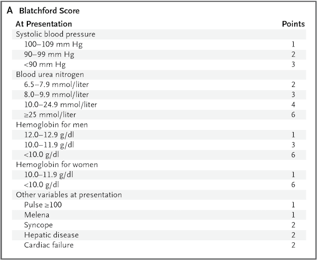
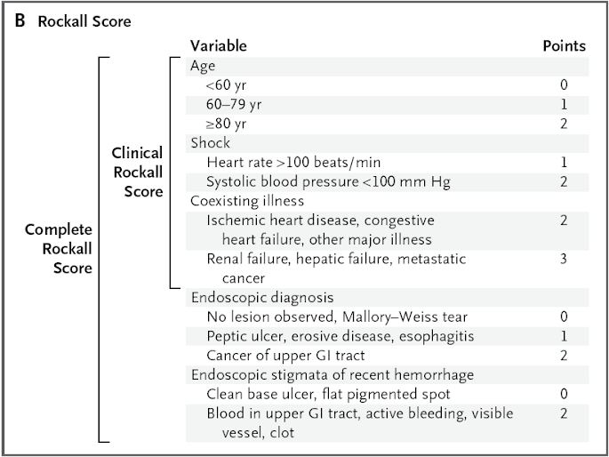
    8. Perform risk stratification; consider the use of a scoring tool (e.g., Blatchford score or clinical Rockall score) before and (e.g., complete Rockall score) after endoscopy.

    Predictors of failure of endoscopic treatment
    • history of peptic ulcer disease
    • previous ulcer bleeding
    • presence of shock at presentation
    • active bleeding during endoscopy
    • large ulcers (>2 cm in diameter)
    • large underlying bleeding vessel (2 mm in diameter)
    • ulcers located on the lesser curve of the stomach or on the posterior or superior duodenal bulb
    Repeat endoscopy may be considered on recurrent bleeding or if there is uncertainty regarding the effectiveness of hemostasis during the initial treatment.

    Surgery remains an effective and safe approach for treating uncontrolled bleeding (i.e., those in whom hemodynamic stabilization cannot be achieved through intravascular volume replacement using crystalloid fluids or blood products) or patients who may not tolerate recurrent or worsening bleeding.

    Angiography with TAE is reserved for patients in whom endoscopic therapy has failed, especially if such patients are high-risk surgical candidates.

    2010年3月8日 星期一

    肝硬化 食道靜脈曲張出血的處置


    Management of Varices and Variceal Hemorrhage in Cirrhosis N Engl J Med March 4, 2010;362:823-32 Purpose of Therapy
    1. Primary prophylaxis: prevent a first episode of variceal hemorrhage
    2. Treatment of the acute bleeding episode
    3. Secondary prophylaxis: prevention of recurrent variceal hemorrhage
    Risk Stratification for Patients with Portal Hypertension 1. Esophagogastroduodenoscopy
    • to evaluate the presence of varices, red wale marks, and variceal size
    2. HVPG (hepatic venous pressure gradient)
    • Portal hypertension is clinically significant when HVPG >10 mmHg, this pressure is the strongest predictor of the development of varices, clinical decompensation, and HCC.
    • In patients with variceal hemorrhage, HVPG >20 mmHg (measured within 24 hours after admission) is the best predictor of a poor outcome.
    3. Child-Turcotte-Pugh classification system
      Treatment of Acute Variceal Hemorrhage Standard Therapy
      1. Combination of vasoconstrictor (terlipressin, somatostatin, or octreotide, administered from the time of admission and maintained for 2 to 5 days) and endoscopic therapy (preferably endoscopic variceal ligation, performed at diagnostic endoscopy <12 hours after admission), together with short-term prophylactic antibiotics (either norfloxacin or ceftriaxone).
      2. Placement of a TIPS is currently considered a salvage therapy for the 10 to 20% of patients in whom standard medical therapy fails.
      3. Endoscopic variceal obturation is the best endoscopic technique to control acute hemorrhage and the TIPS is more effective than variceal obturation in preventing recurrent hemorrhage.