顯示具有 Cardiology 標籤的文章。 顯示所有文章

2018年11月8日 星期四

Management of Heart Failure in ED


Management of Heart Failure in the ED Setting:
An Evidence-Based Review of the Literature

J Emerg Med, 2018 Sep 26.
doi: 10.1016/j.jemermed.2018.08.002

Management of Heart Failure in ED

hemodynamic stabilization and symptom relief
ABC

Mild AHF Exacerbation with Systemic Overload
  • Diuretics (bolus or continuous infusion and high- vs. low-dose)
  • Ultrafiltration for refractory AHF
  • Morphine is not recommended

Hypertensive AHF with Pulmonary Edema
  • NIPPV
  • Nitrates (SL, IV)
  • Diuretics

Hypotensive AHF with Cardiogenic Shock
  • Inotropic agents (norepinephrine ± dobutamine)
  • A small fluid bolus 250–500 mL
  • NIPPV or ETT
  • Mechanical circulatory support: IABP, VAD, V-A ECMO.

High-Output Heart Failure
  • Treat underlying etiology

Disposition
  • 80% admission
  • Observation units

Heart Failure Risk Scores

Af with AHF
  • Cardioversion 
  • Digoxin

2016年4月26日 星期二

Point-of-Care Cardiac Ultrasound


Image-Based Resuscitation of the Hypotensive Patient with Cardiac Ultrasound: An Evidence-Based Review

J Trauma Acute Care Surg. 2016;80:511-518.


Focused Cardiac Ultrasound (FoCUS) 只需簡單的 echo 機, 在 bedsides 即可操作。任何臨床醫師, EP, PA, R, 甚至醫學生都可學習上手,為所有急重症照護者必備的臨床技能。

  • 檢查重點:①心臟收縮功能 ②RV, LV 大小 ③有無 pericardial effusion
  • M mode:① 檢查 cardiac wall 的收縮 ②測量 vena cava 的大小
  • 臨床應用:①鑑別診斷休克的原因 ②決定是否需要灌水
  • Hypovolemia:① empty heart: kissing LV wall ② flat IVC: : IVC collapses >50% during the respiratory cycle
  • Pulmonary embolism: RV enlargement. paradoxic septal movement. D-sign: LV collapse
  • Cardiac tamponade: pericardial effusion + right heart collapse
  • Lung Ultrasound: 看 A line 和 B line (一橫一縱)
  • Interstitial lung edema: lung rockets, B+ lines
  • Pneumothorax: stratosphere sign (B line 可排除此診斷)


【延伸閱讀】
  • RUSH Exam: Rapid Ultrasound in SHock. Emerg Med Clin N Am 28 (2010) 29–56
  • BLUE Protocol: Diagnosis of Acute Respiratory Failure. Chest 2008;134;117-125
  • FALLS Protocol: Fluid administration limited by lung sonography. Expert Rev
    Respir Med 2012; 6: 155-62.
  • C.A.U.S.E.: Cardiac arrest ultrasound exam. Resuscitation (2008) 76, 198—206


2015年6月30日 星期二

Evaluation and Management of Acute Aortic Dissection: ACEP Policy


ACEP Clinical Policy

Evaluation and Management of Adult Patients With Suspected Acute Nontraumatic Thoracic Aortic Dissection

Ann Emerg Med. 2015;65:32-42


Q1. 是否有 clinical decision rules 可以識別 very low risk 的胸主動脈剝離患者?
A: 不要單獨利用目前現有的 clinical decision rules 來識別 very low risk 的胸主動脈剝離患者。

Q2. D-dimer negative 是否可以識別 very low risk 的胸主動脈剝離患者?
A: 不要僅依靠 D-dimer negative 來排除胸主動脈剝離的診斷。

Q3. 與 TEE 和 MRA 相較,CTA 是否夠精確來排除胸主動脈剝離的診斷?
A: 急診可用 CTA 來診斷胸主動脈剝離。其精確性與 TEE, MRA 相近。

Q4. Bedside 心臟超音波異常,可否可確診胸主動脈剝離?
A: 不要依靠 bedside 心臟超音波的異常,來確診胸主動脈剝離。

Q5. 控制心律和血壓,是否可降低胸主動脈剝離的 morbidity or mortality?
A: 胸主動脈剝離患者必需控制心律和血壓。但目前並無明確的目標值,可證實降低胸主動脈剝離的 morbidity or mortality.

2014年12月30日 星期二

2014 AHA/ACC/HRS Atrial Fibrillation Guidelines


2014 AHA/ACC/HRS Guideline for the Management of Patients With Atrial Fibrillation: Executive Summary 
Circulation 2014;130:2071-2104.

Thrombo-embolic Risk and Treatment

  • With prior stroke, TIA, or CHA2DS2-VASc score ≥2, oral anticoagulants recommended. Options include:
Warfarin
– Dabigatran, rivaroxaban, or apixaban
  • With nonvalvular AF and CHA2DS2-VASc score of 0, it is reasonable to omit antithrombotic therapy
  • With nonvalvular AF and a CHA2DS2-VASc score of 1, no antithrombotic therapy or treatment with oral anticoagulant or aspirin may be considered
  • After coronary revascularization in patients with CHA2DS2-VASc score ≥2, it may be reasonable to use clopidogrel concurrently with oral anticoagulants but without aspirin

    Rate Control

    • Control ventricular rate using a beta blocker or non-DHP CCBs for paroxysmal, persistent, or permanent AF
    • In hemodynamically unstable patients, electrical cardioversion is indicated
    • A heart rate control (resting heart rate < 80) strategy is reasonable for symptomatic management of AF
    • IV amiodarone can be useful for rate control in critically ill patients without pre-excitation
    • A lenient rate control strategy (resting heart rate < 110) may be reasonable when patients remain asymptomatic and LV systolic function is preserved
    • Non-DHP CCBs should not be used in decompensated HF

    Rhythm Control  

    • With AF or atrial flutter for >48 h or unknown duration, requiring immediate cardioversion, anticoagulate as soon as possible and continue for at least 4 wk
    • DC cardioversion is recommended for AF or atrial flutter with RVR, that does not respond to pharmacological therapies and contributes to ongoing myocardial ischemia, hypotension, or HF
    • Pharmacological cardioversion: Flecainide, dofetilide, propafenone, ibutilide and amiodarone are useful for cardioversion of AF or atrial flutter


      Specific Patient Groups

      AF complicating ACS
      • Urgent cardioversion of new-onset AF in the setting of ACS is recommended for patients with hemodynamic compromise, ongoing ischemia, or inadequate rate control
      • With ACS and AF with CHA2DS2-VASc score ≥2, anticoagulation with warfarin is recommended unless contraindicated
      • Amiodarone or digoxin may be considered to slow RVR with ACS and AF and severe LV dysfunction and HF or hemodynamic instability
      WPW and pre-excitation syndromes
      • Cardioversion is recommended for patients with AF, WPW syndrome, and RVR who are hemodynamically compromised
      • IV amiodarone, adenosine, digoxin, or non-DHP CCBs in patients with WPW syndrome who have pre-excited AF is potentially harmful
      Heart failure
      • A beta blocker or non-DHP CCB is recommended for persistent or permanent AF in patients with HFpEF
      • IV digoxin or amiodarone is recommended to control heart rate acutely
      • Digoxin is effective to control resting heart rate with HFrEF
      • Amiodarone may be considered when heart rate cannot be controlled with a beta blocker (or a non-DHP CCB with HFpEF) or digoxin, alone or in combination

      【摘要】Atrial Fibrillation 處置

      • CHA2DS2-VASc score ≥2,給 warfarin
      • CHA2DS2-VASc score 0,不需 warfarin
      • 血液動力學不穩定、心肌缺氧症狀、低血壓→Cardioversion
      • Rate control:beta blocker 或 verapamil or diltiazem 無效,給 amiodarone
      • Rate control:有症狀→HR 控制在 80以下;無症狀→HR 控制在 110以下
      • Rhythm control: propafenone, amiodarone
      • Rhythm control:AF >48hrs,整流後立刻給抗凝劑
      • ACS or HF: IV digoxin or amiodarone 
      • WPW syndrome with AF→Cardioversion!禁用 amiodarone, adenosine, digoxin, beta blocker, non-DHP CCBs

      2014年10月15日 星期三

      Non–ST-Elevation–ACS 2014 Guidelines


      Non–ST-Elevation–ACS 2014 Guidelines

      Circulation. published online September 23, 2014


      Prognosis: Early Risk Stratification
      • Perform rapid determination of likelihood of ACS, including a 12-lead ECG within 10 min of arrival at ED (Class I)
      • Perform serial ECGs at 15- to 30-min intervals during the first hour in symptomatic patients with initial non-diagnostic ECG (Class I)
      • Measure cardiac troponin (cTnI or cTnT) in all patients with symptoms consistent with ACS (Class I)
      • Measure serial cardiac troponin at presentation and 3–6 h after symptom onset in all patients with symptoms consistent with ACS (Class I)
      Biomarkers: Diagnosis
      • Measure cardiac-specific troponin (troponin I or T) at presentation and 3─6 h after symptom onset in all patients with suspected ACS to identify pattern of values (Class I)
      • Obtain additional troponin levels beyond 6 h in patients with initial normal serial troponins with ECG changes and/or intermediate/high risk clinical features (Class I)
      • With contemporary troponin assays, CK-MB and myoglobin are not useful for diagnosis of ACS (Class III)

      Early Hospital Care
        Oxygen
      • Administer supplemental oxygen only with SpO2 < 90,  respiratory distress, or other high-risk features for hypoxemia (Class I)
        Nitrates
      • Administer sublingual NTG every 5 min × 3 for continuing ischemic pain and then assess need for IV NTG (Class I)
      • Administer IV NTG for persistent ischemia, HF, or hypertension (Class I)
        Beta-adrenergic blockers
      • Initiate oral beta blockers within the first 24 h in the absence of HF, low-output state, risk for cardiogenic shock, or other contraindications to beta blockade (Class I)
      • Use of sustained-release metoprolol succinate, carvedilol, or bisoprolol is recommended for beta-blocker therapy with concomitant NSTE-ACS, stabilized HF, and reduced systolic function (Class I)

      Anticoagulant Therapy
        Aspirin
      • Non–enteric-coated aspirin (162 mg–325 mg) to all patients promptly after presentation and maintenance dose (81 mg/d–162 mg/d) continued indefinitely (Class I)
        P2Y12 inhibitors
      • P2Y12 inhibitors in addition to aspirin should be administered for up to 12 mo to all patients with NSTE-ACS without contraindications who are treated with either an early invasive or ischemia-guided strategy (Class I)
      Clopidogrel (Plavix) 300-mg or 600-mg loading dose, then 75 mg QD
      Ticagrelor (Brilinta) 180-mg loading dose, then 90 mg BID
      • Ticagrelor in preference to clopidogrel for patients treated with an early invasive or ischemia-guided strategy (Class IIa)
        GP IIb/IIIa inhibitors
      • GP IIb/IIIa inhibitor [Eptifibatide (Integrilin) or tirofiban (Aggrastat)] in patients treated with an early invasive strategy and dual anti-platelet therapy (DAPT) with intermediate/high-risk features (e.g., positive troponin) (Class IIb)
        Parenteral anticoagulant therapy
      • SC enoxaparin
      • Bivalirudin
      • SC Fondaparinux
      • IV Heparin 

        Ischemia-Guided Strategy Versus Early Invasive Strategies
        1. Immediate invasive strategy (within 2 h)
        • Refractory angina
        • Signs or symptoms of HF or new or worsening MR
        • Hemodynamic instability
        • Recurrent angina or ischemia at rest or with low-level activities despite intensive medical therapy
        • Sustained VT or VF
        2. Ischemia-guided strategy
        3. Early invasive strategy (within 24 h)
        4. Delayed invasive strategy (within 25-72 h)


        Ten Points to Remember About NSTE-ACS

        1. Door to ECG < 10min
        2. 有症狀的病人,在第一小時,每15到30分鐘可重覆做ECG
        3. Troponin 症狀開始要驗,3到6小時間要追蹤,症狀超過6小時後也要追蹤
        4. 可以驗 Troponin 時,就不需同時驗 CK-MB,但可考慮驗 BNP
        5. SpO2 < 90%、呼吸窘迫或可能會低血氧的病人需給 O2
        6. Ticagrelor (Brilinta) 優於 Clopidrogel (Plavix)
        7. 低風險病患安排 CV OPD F/U, 要給 aspirin, NTG & beta-blocker
        8. NSTE-ACS 的處置由 Initial Invasive versus Conservative Strategy 改為 Ischemia-Guided Strategy versus Early Invasive Strategies
        9. PCI 分成 Immediate invasive (< 2hr)、Early invasive (< 24hr) 和 Delayed invasive (25-72 hr) 
        10. 頑固性心絞痛 ,心臟衰竭或二尖瓣脫垂症狀,血液動力學不穩定,休息時仍反覆心絞痛且藥物治療無效,sustained VT or VF, 建議 2小時內做 PCI

        2012年10月21日 星期日

        Supraventricular Tachycardia: Evaluation and Initial Treatment


        Evaluation and Initial Treatment of Supraventricular Tachycardia

        N Engl J Med 2012;367:1438-48.


        Differential Diagnosis of Supraventricular Tachycardias (SVT)
        • The initial differential diagnosis of SVT should focus on the ventricular response characteristics of regularity, rate, and rapidity of onset, not on the atrial depolarization from the ECG.
        • The regular SVT include sinus tachycardia, atrial flutter, AV nodal reentrant tachycardia, AV reciprocating tachycardia, and atrial tachycardia.
        • The irregular SVT are atrial fibrillation, atrial flutter with variable AV block, and multifocal atrial tachycardia; multiple atrial premature contractions can cause a similar presentation.
        • Sudden onset and termination are characteristic of acute atrial fibrillation and atrial flutter, AV nodal reentrant tachycardia, AV reciprocating tachycardia, and atrial tachycardia.
        • Gradual onset and recession occur with sinus tachycardia, chronic atrial fibrillation and atrial flutter, multifocal atrial tachycardia, and atrial premature contractions.
        • Adenosine blocks the AV node and is useful in distinguishing among SVT but should not be given in the case of irregular wide-complex tachycardias, since it may render these rhythms unstable.
        • After administration of adenosine, slowing of the heart rate is consistent with a diagnosis of sinus tachycardia, atrial tachycardia, atrial fibrillation, or atrial flutter, whereas termination of tachycardia points to AV nodal reentrant tachycardia, AV reciprocating tachycardia, and some atrial tachycardias.

        Differential Diagnosis and Treatment of Narrow-Complex Tachycardias


        Differential Diagnosis and Treatment of Wide-Complex Tachycardias


        101年 急診專科醫師考題


        關於 wide QRS tachycardia 的敘述,下列何者錯誤?
        1. 若有 ventriculo - atrial dissociation,表示為 ventricular origin
        2. 若為 polymorphic ventricular tachycardia 且先前EKG 有 QTc prolongation 或鎂離子濃度偏低的情形,可考慮直接給予 MgSO4
        3. 若有高度一致性之 QRS complex 但規律性極不規則,加上間歇出現 narrow QRS complexes,應考慮為 atrial fibrillation 合併 WPW syndrome
        4. 若血行動力學不穩定,則宜以同步電擊治療
        5. 若血行動力學穩定,不宜使用adenosine 治療

        2012年8月28日 星期二

        Acute Limb Ischemia


        Acute Limb Ischemia
        N Engl J Med 2012;366:2198-206

        Symptoms
        • Pain
        • Paresthesia
        • Weakness or paralysis
        Signs
        • Absent pulses
        • Pallor
        • Cool skin
        • Decreased sensation
        • Decreased strength
        • Limb blood pressure
        Potential Causes
        • Thrombosis of artery or bypass graft
        • Embolism from heart or proximal vessel
        • Dissection
        • Trauma

        Stages of Acute Limb Ischemia


        Treatment
        • Endovascular Revascularization
        • Surgical Revascularization
        • Catheter-directed thrombolysis has the best results in patients with a viable or marginally threatened limb, recent occlusion (no more than 2 weeks’ duration), thrombosis of a synthetic graft or an occluded stent, and at least one identifiable distal runoff vessel.
        • Surgical revascularization is generally preferred for patients with an immediately threatened limb or with symptoms of occlusion for more than 2 weeks.


        Algorithm for the Diagnosis and Treatment of Acute Limb Ischemia


        Conclusions and Recommendations
        • Heparin should be administered as soon as the diagnosis has been made. 
        • In a patient with a viable or marginally threatened limb, imaging studies (duplex ultrasonography, CTA, or MRA) can be obtained to guide therapeutic decisions.
        • In a patient with an immediately threatened limb, such as the patient described in the vignette, emergency angiography followed by catheter-based thrombolysis or thrombectomy or open surgical revascularization is indicated to restore blood flow and preserve limb viability.

        2012年7月12日 星期四

        New Concepts in the Assessment of Syncope


        New Concepts in the Assessment of Syncope

        J Am Coll Cardiol 2012;59:1583–91

        Definition

        Transient loss of consciousness (TLOC) or faint are generic terms that encompass all disorders characterized by transient, self-limited, nontraumatic loss of consciousness.

        Prognosis

        The outcome in patients with syncope is often related to the severity of the underlying disease rather than the syncopal event itself.

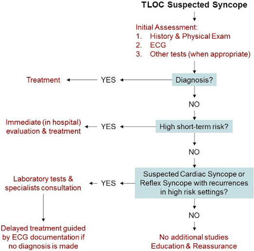
        Classification of Syncope According to Etiology [Modified by ESC (European Society of Cardiology) Guidelines] Versus Classification According to Mechanism [Modified by ISSUE (International Study on Syncope of Uncertain Etiology) Classification]


        The Diagnostic Algorithm of a Patient Presenting With TLOC of Suspected Syncopal Nature


        Short-Term High-Risk Criteria That Require Prompt Hospitalization or Early Intensive Evaluation


        ESC Guidelines
        • Severe structural or coronary artery disease (heart failure, low ejection fraction, or previous myocardial infarction)
        • ECG features suggesting arrhythmic syncope (nonsustained ventricular tachycardia, bifascicular block, inadequate sinus bradycardia ( 50 < bpm) or sinoatrial block, pre-excited QRS complex, ECG findings suggesting an inherited disease)
        • Clinical features suggesting arrhythmic syncope (syncope during exertion or supine position, palpitations at the time of syncope, family history of sudden cardiac death)
        • Important comorbidities:
        • Severe anemia
        • Electrolyte disturbance

        Canadian Cardiovascular Society Position Paper
        • Heart failure and history of cardiac disease (ischemic, arrhythmic, obstructive, valvular)
        • Abnormal ECG (any bradyarrhythmia, tachyarrhythmia, or conduction disease; new ischemia or old infarct)
        • Hypotension (SBP < 90 mmHg)
        • Minor risk factors deserving urgent specialist assessment: age > 60 years, dyspnea, anemia (hematocrit < 30%), hypertension, cerebrovascular disease, family history of sudden death 50 years, syncope while supine, syncope during exercise, syncope with no prodromal symptoms

        2012年4月6日 星期五

        Management of IlioFemoral Deep Vein Thrombosis

        Management of Massive & Submassive Pulmonary Embolism, Iliofemoral Deep Vein Thrombosis, and Chronic Thromboembolic Pulmonary Hypertension

        Circulation. 2011;123:1788-1830


        Initial Anticoagulant Therapy

        • UFH: initial bolus IV 80 U/kg followed by continuous infusion 18 U/kg‧h
        • LMWH: Enoxaparin 1 mg/kg SC BID or 1.5 mg/kg QD
        • Fondaparinux SC QD
        5 mg for patients weighing <50 kg
        7.5 mg for patients weighing 50 to 100 kg
        10 mg for patients weighing >100 kg
        • Adult patients with IFDVT who receive oral warfarin as first-line long-term anticoagulation therapy should have warfarin overlapped with initial anticoagulation therapy for a minimum of 5 days and until the INR is >2.0 for at least 24 hours, and then targeted to an INR of 2.0 to 3.0

        Recommendations for Endovascular Thrombolysis and Surgical Venous Thrombectomy

        Catheter-directed thrombolysis (CDT) or pharmacomechanical CDT should be given to patients with IFDVT associated with limb-threatening circulatory compromise (ie, phlegmasia cerulea dolens)

        2012年3月31日 星期六

        Management of Pulmonary Embolism

        Management of Massive and Submassive Pulmonary Embolism, Iliofemoral Deep Vein Thrombosis, and Chronic Thromboembolic Pulmonary Hypertension

        Circulation 2011, 123:1788-1830


        Definition

        I.   Massive PE

        Acute PE with with at least 1 of the following:
        1.  Sustained hypotension
        SBP <90 mmHg for at least 15 minutes or requiring inotropic support, not due to a cause other than PE, such as arrhythmia, hypovolemia, sepsis, or LV dysfunction
        2. Pulselessness
        3. Persistent profound bradycardia
        heart rate <40 bpm with signs or symptoms of shock

        II.  Submassive PE

        Acute PE without systemic hypotension (SBP >90 mm Hg) but with either RV dysfunction or myocardial necrosis.
        1. RV dysfunction means the presence of at least 1 of the following:
        • Echo: RV dilation (apical 4-chamber RV diameter divided by LV diameter >0.9), or RV systolic dysfunction
        • CT: RV dilation (4-chamber RV diameter divided by LV diameter > 0.9)
        • BNP > 90 pg/mL or N-terminal pro-BNP > 500 pg/mL
        • ECG changes: New complete or incomplete RBBB, anteroseptal ST elevation or depression, or anteroseptal T-wave inversion
        2. Myocardial necrosis is defined as either of the following:
        • Troponin I > 0.4 ng/mL, or Troponin T > 0.1 ng/mL

        I. Low-Risk PE

        Acute PE and the absence of the clinical markers of adverse prognosis that define massive or submassive PE.

        Therapy


        Recommendations for Embolectomy

        • for patients with massive PE and contraindications to fibrinolysis
        • for patients with massive PE who remain unstable after receiving fibrinolysis
        • for patients with submassive acute PE judged to have clinical evidence of adverse prognosis (new hemodynamic instability, worsening respiratory failure, severe RV dysfunction, or major myocardial necrosis)
        • Not recommended for patients with low-risk PE or submassive acute PE with minor RV dysfunction, minor myocardial necrosis, and no clinical worsening

        2012年1月21日 星期六

        How to Interpret Elevated Cardiac Troponin Levels

        How to Interpret Elevated Cardiac Troponin Levels
        Circulation 2011;124:2350-2354.

        Evolution of the cardiac troponin assays and their diagnostic cutoffs

        The 99th Percentile Rule
        In the presence of a clinical history suggestive of ACS, the following is considered indicative of myocardial necrosis consistent with myocardial infarction: maximal concentration of cTn exceeding the 99th percentile of values for a reference control group on at least one occasion during the first 24 hours after the clinical event.


        Causes of Elevated Plasma cTn Other Than ACS
        Cardiac Causes
        • Cardiac contusion resulting from trauma
        • Cardiac surgery
        • Cardioversion
        • Endomyocardial biopsy
        • Acute and chronic heart failure
        • Aortic dissection
        • Aortic valve disease
        • Hypertrophic cardiomyopathy
        • Tachyarrhythmia
        • Bradyarrhythmia, heart block
        • Apical ballooning syndrome
        • Post–PCI
        • Rhabdomyolysis with myocyte necrosis
        • Myocarditis or endocarditis/pericarditis
        Noncardiac Causes
        • Pulmonary embolism
        • Severe pulmonary hypertension
        • Renal failure
        • Stroke, SAH
        • Infiltrative diseases, eg, amyloidosis
        • Cardiotoxic drugs
        • Critical illness
        • Sepsis
        • Extensive burns
        • Extreme exertion

        Serial Troponin Testing
        Collecting a second specimen for cTn testing within 2 to 3 hours from the collection of the blood sample at presentation to help confirm the diagnosis of MI.

        Troponin kinetics in the index cases

        2011年12月19日 星期一

        2010 Guidelines on Thoracic Aortic Disease

        2010 ACCF/AHA/AATS/ACR/ASA/SCA/SCAI/SIR/STS/SVM Guidelines for the Diagnosis and Management of Patients With Thoracic Aortic Disease

        Circulation 2010;121;e266-e369
        Recommendations for Aortic Imaging Techniques
        • For CT or MRI, the external diameter should be measured perpendicular to the axis of blood flow.
        • For echocardiography, the internal diameter should be measured perpendicular to the axis of blood flow.
        • The finding of aortic dissection, aneurysm, traumatic injury and/or aortic rupture should be immediately communicated to the referring physician.

        Aortic Imaging Reports
        • The location at which the aorta is abnormal.
        • The maximum diameter of any dilatation, measured from the external wall of the aorta, perpendicular to the axis of flow, and the length of the aorta that is abnormal.
        • For patients with presumed or documented genetic syndromes at risk for aortic root disease measurements of aortic valve, sinuses of Valsalva, sinotubular junction, and ascending aorta.
        • The presence of internal filling defects consistent with thrombus or atheroma.
        • The presence of intramural hematoma (IMH), penetrating atherosclerotic ulcer (PAU), and calcification.
        • Extension of aortic abnormality into branch vessels, including dissection and aneurysm, and secondary evidence of end-organ injury (eg, renal or bowel
          hypoperfusion).
        • Evidence of aortic rupture, including periaortic and mediastinal hematoma, pericardial and pleural fluid, and contrast extravasation from the aortic lumen.
        • When a prior examination is available, direct image to image comparison to determine if there has been any increase in diameter.

        High Risk Conditions
        • Marfan Syndrome
        • Connective tissue disease
        • Family history of aortic disease
        • Known aortic valve disease
        • Recent aortic manipulation (surgical or catheter-based)
        • Known thoracic aortic aneurysm
        • Genetic conditions that predispose to AoD

        High Risk Pain Features
        Chest, back, or abdominal pain features described as pain that:
        • is abrupt or instantaneous in onset.
        • is severe in intensity.
        • has a ripping, tearing, stabbing, or sharp quality.

        High Risk Examination Features
        • Pulse deficit
        • Systolic BP limb differential > 20mm Hg
        • Focal neurologic deficit
        • Murmur of aortic regurgitation (new or not known to be old and in conjunction with pain)

        Initial Management
        • IV β-blockade should be initiated and titrated to a target heart rate < 60 bpm non-dihydropyridine Ca channel-blockade should be used as an alternative for rate control if contraindications to β-blockade, If SBP > 120mmHg after heart rate control has been obtained, then ACEIs and/or other vasodilators should be administered to further reduce BP that maintains adequate end-organ perfusion.
        • β-blockers should be used cautiously in the setting of acute aortic regurgitation because they will block the compensatory tachycardia.
        • Vasodilator therapy should not be initiated prior to rate control so as to avoid associated reflex tachycardia that may increase aortic wall stress, leading to propagation or expansion of a thoracic aortic dissection.

        AoD Evaluation Pathway

        Acute AoD Management Pathway

        Acute Surgical Management Pathway for AoD

          2011年3月3日 星期四

          2009 ACCF/AHA Heart Failure Guidelines


          2009 Focused Update: ACCF/AHA Guidelines for the Diagnosis and Management of Heart Failure in Adults

          J. Am. Coll. Cardiol. April 14, 2009; 53;1343-1382

          Circulation. April 14, 2009;119;1977-2016

          Initial Clinical Assessment
          • Coronary arteriography should be performed in HF who have angina or significant ischemia unless the pt is not eligible for revascularization.
          • BNP or NT-proNBP can be useful in the urgent care setting in whom the clinical diagnosis of HF is uncertain. 
          • Measurement of BNP can be helpful in risk stratification.
          Patients with Reduced Left Ventricular Ejection Fraction
          • Diuretics and salt restriction
          • ACE inhibitors or angiotensin II receptor blockers
          • beta blockers 
          • Implantable Cardioverter-Defibrillatoras (ICD)
          • ICD is recommended in pts with of HF and reduced LVEF who have a history of cardiac arrest, VF, or hemodynamically destabilizing VT.
          • ICD is recommended for primary prevention of SCD to reduce total mortality in pts with non-ischemic DCM or IHD > 40 days post-MI, LVEF ≤ 35%, NYHA Fc II or III while receiving chronic optimal medical therapy, and who have expectation of survival with good functional status > 1 year.
          • Resynchronization Therapy
          • Digitalis
          • Hydralazine and Nitrate Combination
          • Positive Inotropic Drugs 

          Recommendations for the Hospitalized Patient (New Recommendations)

          Diagnosis of HF

          Clinicians should determine the following:
          • adequacy of systemic perfusion
          • volume status
          • contribution of precipitating factors and/or co-morbidities
          • if the heart failure is new onset or an exacerbation of chronic disease
          • whether it is associated with preserved normal or reduced ejection fraction
          CXR, ECG, and echocardiography are key tests in this assessment
          Patients Being Evaluated for Dyspnea
          • BNP or NT-proBNP should be measured in pts being evaluated for dyspnea in which the contribution of HF is not known.
          • Precipitating Factors for Acute HF
          • ACS/coronary ischemia
          • severe hypertension
          • atrial and ventricular arrhythmias
          • infections
          • pulmonary emboli
          • renal failure
          • medical or dietary non-compliance
          • Oxygen Therapy and Rapid Intervention 
          • Intravenous Loop Diuretics
          • Monitoring Fluid Intake and Output
          • Intensifying the Diuretic Regimen
          • Preserving End-Organ Performance
          In pts with clinical evidence of hypotension associated with hypoperfusion and obvious evidence of elevated cardiac filling pressures (e.g., elevated jugular venous pressure; elevated PAWP), IV inotropic or vasopressor drugs should be administered to maintain systemic perfusion and preserve end-organ performance while more definitive therapy is considered.
          • ACE inhibitors or ARBs and beta-blocker therapy
          • Urgent Cardiac Catheterization and Revascularization
          When pts present with acute HF and known or suspected acute myocardial ischemia due to occlusive coronary disease, especially when there are signs and symptoms of inadequate systemic perfusion.
          • Severe Symptomatic Fluid Overload
          in the absence of systemic hypotension, vasodilators such as IV NTG, nitroprusside or neseritide can be beneficial.
          • Invasive Hemodynamic Monitoring
          for carefully selected pts with acute HF who have persistent symptoms despite empiric adjustment of standard therapies, and
          • whose fluid status, perfusion, or systemic or pulmonary vascular resistances are uncertain
          • whose SBP remains low, associated with symptoms, despite initial therapy
          • whose renal function is worsening with therapy
          • who require parenteral vasoactive agents
          • who may need consideration for advanced device therapy or transplantation
          相關文章:Acute Heart Failure Syndromes

          2011年2月21日 星期一

          Acute Coronary Syndromes


          Acute Coronary Syndromes 2010 AHA Guidelines for CPR and ECC  Circulation. 2010;122:S787-S817 ACC/AHA 2009 Guidelines for STEMI and PCI Circulation. 2009;120;2271-2306 ACC/AHA 2007 Guidelines for UA/NSTEMI Circulation. 2007;116;e148-e304
          TIMI Risk Score
          • Age ≥ 65 years
          • At least 3 risk factors for CAD (family history of CAD, hypertension, hypercholesterolemia, diabetes, or current smoker)
          • Prior coronary stenosis of ≥ 50%
          • ST-segment deviation on ECG
          • At least 2 anginal events in prior 24 hours
          • Use of aspirin in prior 7 days
          • Elevated serum cardiac biomarkers
          PCI Following ROSC After Cardiac Arrest
          • Patients with OHCA due to VF in the setting of STEMI or new LBBB, emergent angiography with prompt recanalization of the infarct-related artery is recommended (Class I, LOE B).
          • PPCI after ROSC in subjects with arrest of presumed ischemic cardiac etiology may be reasonable, even in the absence of a clearly defined STEMI (Class IIb, LOE B).
          • Appropriate treatment of ACS or STEMI, including PCI or fibrinolysis, should be initiated regardless of coma (Class I, LOE B)
          Management of Arrhythmias
          • Primary VF accounts for the majority of early deaths during AMI. The incidence of primary VF is highest during the first 4 hours after onset of symptoms.
          • Prophylactic antiarrhythmics are not recommended for patients with suspected ACS or MI in the prehospital or ED (Class III, LOE A).
          • Routine IV administration of beta-blockers to patients without hemodynamic or electric contraindications is associated with a reduced incidence of primary VF (Class IIb, LOE C).
          • Low serum potassium has been associated with ventricular arrhythmias. It is prudent clinical practice to maintain serum potassium >4 mEq/L (Class IIB, LOE A).

          2010年11月14日 星期日

          Acute Heart Failure Syndromes


          Acute Heart Failure Syndromes: 
          Emergency Department Presentation, Treatment, and Disposition:
          Current Approaches and Future Aims
          Circulation. Nov 9, 2010;122:1975-1996


          Current Diagnostics
          • The evaluation of possible AHFS in the ED includes history, physical examination, CXR, 12-lead ECG,cardiac troponin I or T, electrolytes, and CBC.
          • The BNP and NT-proBNP have demonstrated diagnostic utility when clinical uncertainty remains after initial history, physical examination, and CXR.
          • Either BNP or NT-proBNP should be measured in patients in whom there is clinical uncertainty concerning the diagnosis of AHFS.
          Optimal NT-proBNP cut-points for the diagnosis or exclusion of AHFS
          • BNP and NT-proBNP single cut point of 300 pg/mL to rule out AHFS
          • 2 cut points to rule in AHFS depending on age:
          <50 years old ( >450 pg/mL)
          >50 years old ( >900pg/mL)
          Am J Cardiol. 2005;95:948–954.
          • Age-independent cutoff of 900 pg/mL
          • Age-stratified approach of 450/900/1800 for patients aged <50/50 to 75/>75 years
          Category
          Optimal cut-point
          Sensitivity  (%)
          Specificity (%)
          PPV (%)
          NPV (%)
          Accuracy (%)
          Confirmatory (‘rule in’) cut-points
          <50 years (n=184)
          450 pg/mL
          97
          93
          76
          99
          94
          50–75 years (n=537)
          900 pg/mL
          90
          82
          83
          88
          85
          >75 years (n=535)
          1800 pg/mL
          85
          73
          92
          55
          83
          Rule in, overall
          90
          84
          88
          66
          85
          Exclusionary (‘rule out’) cut-point
          All patients (n=1256)
          300 pg/mL
          99
          60
          77
          98
          83
          Eur Heart J. 2006;27:330–337
          Am J Cardiol. 2008;101:29–38
          Current Therapy: Heterogeneous Presentations Met With Homogeneous Therapy
            • Regardless of the baseline cardiac pathophysiology, critical presenting features such as hemodynamic status, presence of myocardial ischemia, and renal dysfunction, the current goals of ED therapy are to relieve congestion, balance hemodynamics, achieve euvolemia.
            • Initial stabilization focuses on determining whether the patient requires ventilatory support.
            • Diuretics and vasodilators are frequently used in the treatment of AHFS with congestion and normal or elevated blood pressure.
            • Hypertension may appear to be the most acutely ill, but aggressive blood pressure management often results in rapid resolution of symptoms. 
            ED Disposition
            • 80% of patients who present to the ED with AHFS are hospitalized.
            Predictors of Adverse Events
            • Elevated BUN or creatinine 
            • Hyponatremia 
            • Ischemic ECG changes 
            • Elevated BNP levels 
            • Elevated troponins 
            • Low systolic blood pressure 
            Morbidity and Mortality in Hospitalized Patients with AHFS
            • The average risk of death during hospital admission for AHFS is apprximately 4%. 
            • Patients requiring the use of inotropic agents had a mortality rate of 12% to 13%. 
            • Traditional HF medical therapy including ACE inhibitors, ARB, β-blockers, and selective aldosterone receptor antagonists. Early initiation of this therapy, before hospital discharge, with appropriate titration, improves symptoms, reduces hospitalizations, and saves lives. 
            Patient Characterization
            The European Society of Cardiology classification:
            1. Worsening or decompensated chronic HF 
            2. Cardiogenic pulmonary edema 
            3. Hypertensive AHFS 
            4. Cardiogenic shock 
            5. Isolated right HF 
            6. AHFS with ACS
            • Nitrates might be used in higher relative doses to diuretics in the hypertensive profile Ultrafiltration could be usedin the diuretic-resistant patient 
            • Inotropic agents should be considered in the rarer cases of advanced/low-output HF 
            • Hypotension and tachycardia should be avoided, especially in patients with coronary artery disease 
            ACC/AHA Stages of Heart Failure
            • A: At high risk for HF but without structural heart disease or symptoms of HF 
            • B: Structural heart disease but without signs or symptoms of HF 
            • C: Structural heart disease with prior or current symptoms of HF 
            • D: Refractory HF requiring specialized interventions
            Risk Stratification: Low-Risk, Not High-Risk Markers Are Necessary
            • Hypotension, hyponatremia, renal dysfunction, increased troponin levels, and elevated BNP all portend a poor prognosis. 
            • The absence of high-risk markers does not define a low-risk patient.
            • Data suggest that EP would be comfortable discharging a patient if there was a combined overall risk of in-hospital events or 30-day mortality of <2%. 
            第4張 slide 更正:
            • BNP and NT-proBNP can be elevated in sepsis, pulmonary hypertension, older age, renal insufficiency, atrial fibrillation, pulmonary embolism and obesity. 
            • Obesity is associated with disproportionately low BNP levels.

            2010年11月5日 星期五

            ACC/AHA 2009 Guidelines for STEMI & PCI


            ACC/AHA 2009 STEMI/PCI Guidelines 
            ACC/AHA Guidelines for the Management of Patients with ST-Elevation Myocardial Infarction (STEMI) and the ACC/AHA/SCAI Guidelines on Percutaneous Coronary Intervention (PCI)

            J. Am. Coll. Cardiol.  2009;54;2205-2241
            Circulation. 2009;120;2271-2306

            Glycoprotein IIb/IIIa Antagonists in STEMI
            • It is reasonable to start treatment with GP IIb/IIIa antagonists (abciximab, tirofiban and eptifibatide) at the time of primary PCI in selected patients with STEMI
            Thienopyridine in STEMI
            A loading dose of thienopyridine is recommended for STEMI patients for whom PCI is planned.
            Regimens should be one of the following:
            • Clopidogrel at least 300 to 600 mg should be given as early as possible before or at the time of primary or non-primary PCI.
            • Prasugrel 60 mg should be given as soon as possible for primary PCI.
            Parenteral Anticoagulants in STEMI
            • For prior treatment with UFH, additional boluses of UFH should be administered as needed to maintain therapeutic activated clotting time levels, taking into account whether GP IIb/IIIa antagonists have been administered; or
            • Bivalirudin is useful as support for primary PCI with or without prior treatment with heparin.
            Triage and Transfer for PCI in STEMI
            High Risk Definition: 
            • Defined in CARESS-in-AMI as STEMI patients with one or more high-risk features:
            – extensive ST-segment elevation
            – new-onset left BBB
            – previous MI
            – Killip class >2
            – LVEF <35% for inferior MI
            Anterior MI alone with 2 mm or more ST-elevation in 2 or more leads qualifies
            • Defined in TRANSFER-AMI as >2 mm ST-segment elevation in 2 anterior leads or ST elevation at least 1 mm in inferior leads with at least one of the following:
            – SBP <100 mmHg
            – heart rate >100 bpm
            – Killip Class II-III
            – >2 mm of ST-segment depression in the anterior leads
            – >1mm of ST elevation in right-sided lead V4
            相關文章:
            ACC/AHA 2007 Guidelines for UA/NSTEMI

            2010年9月14日 星期二

            ACC/AHA 2007 Guidelines for UA/NSTEMI


            ACC/AHA 2007 Guidelines for the Management of Patients with Unstable Angina/Non-ST-Elevation Myocardial Infarction  Circulation. 2007;116;e148-e304 J. Am. Coll. Cardiol. 2007;50;652-726  Initial Evaluation and Management of UA/NSTEMI  Clinical Assessment
            • 12-lead ECG should be performed and shown to EP as soon as possible, with a goal of within 10 min of ED arrival for all patients with chest discomfort or symptoms suggestive of ACS.
            • If initial ECG is not diagnostic but patient remains symptomatic and there is high clinical suspicion for ACS, serial ECGs, initially at 15- to 30-min intervals, should be performed to detect the potential for development of ST-segment elevation or depression. 
            Early Risk Stratification TIMI Risk Score
            • Age ≥ 65 years
            • At least 3 risk factors for CAD (family history of CAD, hypertension, hypercholesterolemia, diabetes, or current smoker)
            • Prior coronary stenosis of ≥ 50%
            • ST-segment deviation on ECG
            • At least 2 anginal events in prior 24 hours
            • Use of aspirin in prior 7 days
            • Elevated serum cardiac biomarkers 
            GRACE risk score
            Selection of Initial Treatment Strategy Initial Invasive versus Conservative Strategy Invasive
            • Recurrent angina/ischemia at rest with low-level activities despite intensive medical therapy
            • Elevated cardiac biomarkers (TnT or TnI)
            • New/presumably new ST-segment depression
            • Signs/symptoms of HF or new/worsening MR
            • High-risk findings from noninvasive testing
            • Hemodynamic instability
            • Sustained VT 
            • PCI within 6 months 
            • Prior CABG 
            • High risk score (e.g., TIMI, GRACE)
            • Reduced left ventricular function (LVEF < 40%)
            Conservative
            • Low risk score (e.g., TIMI, GRACE)
            • Patient/physician presence in the absence of high-risk features 
            Stepped-Care Approach to Pharmacological Therapy for Musculoskeletal Symptoms with Known Cardiovascular Disease or Risk Factors for Ischemic Heart Disease
               

            ED Management Strategies: Guidelines at a Glance