顯示具有 Trauma 標籤的文章。 顯示所有文章

2017年12月18日 星期一

ATLS® 第10版 更新摘要


ATLS 10th Edition Compendium of Change

Primary Survey
Airway Maintenance with Restriction of Cervical Spine Motion
  • “cervical spine protection” changed to ”Restriction of Cervical Spine Motion
  • RSI changed to ”Drug Assisted Intubation
Breathing and Ventilation 
Circulation with Hemorrhage Control
  • Initial resuscitation: Adult: 1 L isotonic solution; Child < 40 kg: 20 ml/kg
  • Tranexamic acid: 1 g over 10 min within 3 hr, then 1 g over 8 hr
  • Hemorrhagic shock classification table amended: Base excess 

Thoracic Trauma
  • Life threatening chest injury: flail chest out, tracheobrochial injury now in
  • Tension pneumothorax:
    • Needle thoracocentesis
      • 5th  ICS mid-axillary line for adult
      • UNCHANGED 2nd ICS mid-clavicular line for child
    • 28-32 Fr chest drain for hemothorax (not 36-40 Fr)
      • eFAST (extended FAST): seashore, bar code, or stratosphere sign in M mode
    • Aortic rupture management with Beta Blocker (esmolol): goal heart rate < 80 bpm and MAP 60-70 mmHg
    • Algorithm for circulation arrest approach

    Abdominal and Pelvic Trauma
    • Palpation of prostate gland no longer recommended for urethral injury

    Head Trauma
    • Classification: “minor” changed to “mild” head trauma
    • Detailed guidance on SBP management
      • Maintain SBP at ≥ 100 mmHg for patients 50-69 years or at ≥ 110 mmHg for patients 15-49 years or older than 70 years.
    • Anticoagulation reversal guidance

    Spine and Spinal Cord Trauma
    • New myotome diagram
    • Canadian C-Spine Rule (CCR) and NEXUS Criteria


      Musculoskeletal Trauma
      • Highlighting risk factor of bilateral femur fractures

      Thermal Injury

      Paediatric Trauma
      • Pediatric Emergency Care Applied Research Network (PECARN) Criteria for Head CT

        Transfer to Definitive Care
        • Specific mention of avoiding CT in primary hospital
        • SBAR template for communication

        Mobile ATLS: New to this edition

        Royal College Surgeons ATLS course 時程公告:
        2018年 2月   以前,ATLS 第 9 版
        2018年 3月到 5月,ATLS 第 9 版 + 第 10版 重點導讀
        2018年 6月   以後,ATLS 第 10版

        2017年9月7日 星期四

        The European Guideline on Management of Major Bleeding and Coagulopathy Following Trauma: 4E


        The European Guideline on Management of Major Bleeding and Coagulopathy Following Trauma: Fourth Edition

        Rossaint et al. Critical Care (2016) 20:100
        DOI 10.1186/s13054-016-1265-x


        Major Bleeding
        • FAST, CT 找出血
        • Damage control surgery if shock or coagulopathy
        • Damage control resuscitation 直到找到出血來源並控制
        • Restricted volume replacement: target SBP: 80-90 mmHg; severe TBI (GCS ≤8): MAP ≥80 mmHg
        • Tranexamic acid (TXA) as early as possible (< 3hr): loading dose TXA 1 g over 10 min, followed by TXA 1 g over 8 h
        Transfusion
        • Restrictive RBC transfusion: target Hb 7–9 g/dl
        • FFP-RBC ratio >1:2
        • Fibrinogen maintain at 1.5–2 g/l
        • FFP administered to maintain PT and APTT ≤ 1.5 times the normal control
        • Platelet count >100K
        • PCC administered in patients pre-treated with warfarin or direct-acting oral coagulants
        • Off-label use of rFVIIa only if major bleeding and traumatic coagulopathy persist despite standard attempts to control bleeding and best practice use of conventional hemostatic measures.

        2015年4月28日 星期二

        C-Spine Collar Clearance In The Obtunded Adult Blunt Trauma Patient


        Cervical Spine Collar Clearance In The Obtunded Adult Blunt Trauma Patient
        A systematic review and practice management guideline from the Eastern Association for the Surgery of Trauma

        J Trauma Acute Care Surg. 2015;78: 430-441.


        PICO

        • Population: In the obtunded adult blunt trauma patient
        • Intervention: Should cervical collar removal be performed after a negative high-quality C-spine CT result combined with adjunct imaging?
        • Comparator: Should cervical collar removal be performed after a negative high-quality C-spine CT result alone?
        • Outcome:To reduce peri-clearance events, such as new neurologic change (paraplegia, quadriplegia), unstable C-spine injury (subcategories, treated with operation or treated with orthotic), stable C-spine injury (subcategories treated with operation or treated with orthotic), post-clearance imaging, false-negative CT imaging result on re-review, pressure ulcers, and time to cervical collar clearance.

        Inclusion Criteria

        • Adult blunt trauma patients > 16 years
        • C-spine CT with axial thickness < 3 mm
        • Obtunded definition: GCS score < 15, unconscious, intubated, altered mental status, un-reliable examination, distracting injury, intoxication, or not meeting NEXUS guidelines

        RECOMMENDATION

        In obtunded adult blunt trauma patients, we conditionally recommend cervical collar removal after a negative high-quality C-spine CT scan result alone.

        NPV of CT C-spine for an Unstable Injury: 100%
        NPV of CT C-spine for an Stable Injury: 90.6% 


        【摘要】 意識不清傷患之頸圈移除建議 ﹝2015 EAST Guideline﹞

        C-spine CT negative finding 之意識不清傷患,建議視情況移除頸圈

        ﹝Unstable C-Spine Injury NPV: 100%; Stable C-Spine Injury NPV: 90.6%﹞

        2014年4月2日 星期三

        Major Changes In The ATLS 9th Edition


        ATLS® 第9版 重大改變

        Journal of Trauma and Acute Care Surgery
        2013; 72(5): 1363–1366
        ATLS 平均每四年改版一次,ATLS第8版 已經是 2008年,ATLS第9版 於 2012年底出版,新版主要的改變分為三個部分:


        1. 內容的改變
        • Team Training: 在初步評估章節中,強調創傷團隊訓練
        • Airway:
        強調進階呼吸道處置和 videolaryngoscopy
        除了小於一歲的嬰兒外,所有小兒插管可使用 cuffed endotracheal tubes
        • Balanced Resuscitation
        積極輸液 (aggressive resuscitation) 已被刪除,過去標準一開始就給 2L 的輸液改為 1-2 L
        休克傷患強調早期輸血及血小板
        2. 架構的改變
        • Pelvis: 強調骨盆腔為出血來源,並將所有內容移至 abdomen & pelvis 章節
        • 技術站: DPL 和 pericardiocentesis 列為選修。腹部的出血評估如果沒有教 DPL, 就一定要教 FAST
        • 初步評估情境題: 強調老人、鈍挫傷、肋骨骨折處置、骨盆骨折

        3. MyATLS
        原先的 DVD 光碟變成了 MyATLS app
        iOS
        Android

        補充資料:ATLS 9th Edition Compendium of Change
        相關文章:ATLS 8e, The Evidence for Change

        2013年4月16日 星期二

        爆炸傷害的診斷和處置


        Primary blast injury: Update on diagnosis and treatment

        Crit Care Med 2008; 36:[Suppl.]:S311–S317


        Injuries from explosions are traditionally classified into:
        1. Primary blast injuries: injuries due solely to the blast wave
        2. Secondary blast or explosive injury: primarily ballistic trauma resulting from fragmentation wounds from the explosive device or the environment
        3. Tertiary blast or explosive injury: result of displacement of the victim or environmental structures, is largely blunt traumatic injuries
        4. Quaternary explosive injuries: burns, toxins, and radiologic contamination

        Ocular Injury
        • Ophthalmology consultation should be obtained for suspected globe injuries, corneal foreign bodies or abrasions, orbital fractures, retinal detachments, hyphema, intraocular foreign bodies, corneal or eyelid burns, lid lacerations, subconjunctival hemorrhage, or head injuries that involve the orbit or may compromise vision
        Aural Injury
        • Tympanic membrane rupture is the most common primary blast injury
        • Clinicians should make otoscopic examination a routine part of the initial evaluation of explosion injured patients
        Pulmonary Injury
        • Blast lung injury is the most common fatal injury among initial survivors of explosions
        • Clinical diagnosis of blast lung injury is based on the presence of respiratory distress, hypoxia, and “butterfly” or batwing infiltrates.
        CVS Injury
        • Triad of immediate bradycardia, hypotension, and apnea.
        GI Injury
        • Mesenteric or mural hematoma in hemodynamically stable patients without peritoneal signs may be managed with NPO, NG tube decompression, and resuscitation.
        • Massive hemorrhage or obvious hollow viscus perforation should be treated with laparotomy for hemostasis and control of spillage of enteric contents.
        Traumatic Amputations
        • Early tourniquet use
        Brain Injury
        • PE should include a thorough NE to include checking for positive Romberg's sign as well as funduscopy to look for evidence of air emboli.
        • CT scan should be used to search for evidence of blunt head injury and ICH.

        Patient Risk Stratification


        【重點摘要】
        1. 初級爆炸傷害:直接由爆炸波導致的傷害
        2. 次級爆炸傷害:因爆炸物或環境的碎片導致的傷害
        3. 三級爆炸傷害:因傷患或環境的位移導致的傷害,主要是鈍挫傷
        4. 四級爆炸傷害:燒燙傷、毒物或輻射污染

        耳道傷害
        • 鼓膜破裂 是最常見的原發性爆炸傷害
        • 臨床醫生對於所有爆炸受傷的病人,常規性的耳鏡檢查,應列為初步評估的一部分

        肺傷害
        • 爆炸性肺損傷 是爆炸倖存者早期最常見的致命傷害
        • 臨床診斷:呼吸窘迫,缺氧,X光會有“蝴蝶 butterfly” 或 “蝙蝠 batwing” 浸潤

        2012年8月14日 星期二

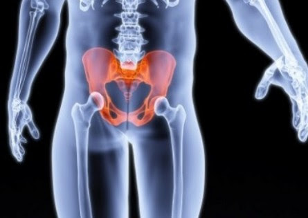
        EAST Pelvic Fracture Guidelines


        Eastern Association for the Surgery of Trauma Practice Management Guidelines for Hemorrhage in Pelvic Fracture—Update and Systematic Review

        J Trauma. 2011;71: 1850–1868


        Six specific questions are addressed regarding the management of pelvic fracture hemorrhage:

        1. Which Patients With Hemodynamically Unstable Pelvic Fractures Warrant Early External Mechanical Stabilization?

          • The use of a pelvic orthotic device (POD) does not seem to limit blood loss in patients with pelvic hemorrhage. Level III recommendation
          • The use of a POD effectively reduces fracture displacement and decreases pelvic volume. Level III recommendation

          • Which Patients Require Emergent Angiography?

            • Patients with pelvic fractures and hemodynamic instability or signs of ongoing bleeding after nonpelvic sources of blood loss have been ruled out should be considered for pelvic angiography/embolization. Level I recommendation
            • Patients with evidence of arterial intravenous contrast extravasation in the pelvis by CT may require pelvic angiography and embolization regardless of hemodynamic status. Level I recommendation
            • Patients with pelvic fractures who have undergone pelvic angiography with or without embolization, who have signs of ongoing bleeding after nonpelvic sources of blood loss have been ruled out, should be considered for repeat pelvic angiography and possible embolization. Level II recommendation
            • Patients older than 60 years with major pelvic fracture (open book, butterfly segment, or vertical shear) should be considered for pelvic angiography without regard for hemodynamic status. Level II recommendation
            • Although fracture pattern or type does not predict arterial injury or need for angiography, anterior fractures are more highly associated with anterior vascular injuries, whereas posterior fractures are more highly associated with posterior vascular injuries. Level III recommendation
            • Pelvic angiography with bilateral embolization seems to be safe with few major complications. Gluteal muscle ischemia/necrosis has been reported in patients with hemodynamic instability and prolonged immobilization or primary trauma to the gluteal region as the possible cause, rather than a direct complication of angioembolization. Level III recommendation
            • Sexual function in males does not seem to be impaired after bilateral internal iliac arterial embolization. Level III recommendation 
             
          • What Is the Best Test to Exclude Intra-Abdominal Bleeding?

            • Focused Assessment with Sonography for Trauma (FAST) is not sensitive enough to exclude intraperitoneal bleeding in the presence of pelvic fracture. Level I recommendation
            • FAST has adequate specificity in patients with unstable vital signs and pelvis fracture to recommend laparotomy to control hemorrhage. Level I recommendation
            • Diagnostic peritoneal tap (DP)/Diagnostic peritoneal lavage (DPL) is the best test to exclude intra-abdominal bleeding in the hemodynamically unstable patient. Level II recommendation 
            • In the hemodynamically stable patient with a pelvic fracture, CT of the abdomen and pelvis with intravenous contrast is recommended to evaluate for intra-abdominal bleeding regardless of FAST results. Level II recommendation
             
            1. Are There Radiologic Findings Which Predict Hemorrhage?

              • Fracture pattern on pelvic X-ray does not single-handedly predict mortality, hemorrhage, or the need for angiography. Level II recommendation
              • Presence/location of hematoma does not predict or exclude the need for angiography and possible embolization. Level II recommendation
              • CT of the pelvis is an excellent screening tool to exclude pelvic hemorrhage. Level II recommendation
              • Absence of contrast extravasation on CT does not always exclude active hemorrhage. Level II recommendation 
              • Pelvic hematoma 500 cc in size has an increased incidence of arterial injury and need for angiography. Level II recommendation
              • Isolated acetabular fractures are as likely to require angiography as pelvic rim fractures. Level III recommendation 
              • If a retrograde urethrocystogram is required, it should be performed after CT with intravenous contrast. Level III recommendation
               
            2. What Is the Role of Noninvasive Temporary External Fixation Devices?

              • TPBs effectively reduce unstable pelvic fractures as well as definitive stabilization and decrease pelvic volume. Level III recommendation
              • TPBs may limit pelvic hemorrhage but do not seem to affect mortality. Level III recommendation
              • TPBs work as well or better than emergent EPF in controlling hemorrhage. Level III recommendation 
               
            3. Which Patients Warrant Retroperitoneal (Preperitoneal) Packing?

              • Retroperitoneal pelvic packing is effective in controlling hemorrhage when used as a salvage technique after angiographic embolization. Level III recommendation
              • Retroperitoneal pelvic packing is effective in controlling hemorrhage when used as part of a multidisciplinary clinical pathway including a POD/C-clamp. Level III recommendation

            2012年5月17日 星期四

            懷孕創傷

            基本觀念

            • 懷孕創傷是兩個受傷病患
            • Save The Mother First!! Save The Fetus If Possible.
            • 母親死亡是外傷造成胎兒死亡最常見的原因
            • 胎盤剝離是造成胎兒死亡的第二常見原因

            孕婦評估

            • 同 ATLS 流程
            • 對母親最適當的急救,就是對胎兒最好的照護。
            • 孕婦儘可能保持左側躺的姿勢。右髖部墊高、長背板左傾 15-30度

            胎兒評估

            • 胎兒評估始於胎心音監測
            • 創傷產婦,胎兒監測最少應監測 四小時
            • 正常胎心音 FHB:120-160 bpm
            • FHB↓ →缺氧
            • 可能原因:母親低血壓、低體溫、呼吸窘迫、胎盤剝離…
            • 孕婦死亡五分鐘內進行剖腹產,胎兒有良好的預後(70% 存活率)
            • 若無胎心音,不必要進行剖腹產,大部分在一週內會自行產出

            何時要做放射影像檢查?

            • 當臨床狀況必須實施診斷性放射影像檢查時,就不應該因考慮胎兒輻射傷害而延誤檢查
            • 風險評估:診斷性放射檢查對母親的好處,遠超過對胎兒造成的傷害

            孕婦創傷急救流程

            摘要

            • ATLS 流程同非懷孕者
            • 先急救母親,再評估胎兒
            • 孕婦可能保持左側躺的姿勢、左傾15-30度可改善母親和胎兒的循環
            • 必要的放射影像檢查勿因胎兒而延誤
            • 懷孕大於20-24周,胎心音監測至少四到六小時


            急診專科醫師 甄審考題


            關於正常懷孕的生理變化,下列敘述何者錯誤?【99急專】
            1. 白血球數目增加,血紅素濃度減少
            2. 全身血量增加,心臟輸出增加
            3. 收縮壓及舒張壓皆降低,但舒張壓降低較多
            4. 呼吸速度變快,潮氣容積增加
            5. 較大的失血量才會出現週邊灌流不足(休克)的徵象

            治療懷孕的外傷病人時,下列敘述何者正確?【96急專】
            1. 一般治療的優先順序(priority)與未懷孕者是不同的
            2. 先治療孕婦的傷勢再對胎兒做早期評估
            3. 面對孕婦與胎兒若兩者均存活則先治療哪一位以當時狀況而定
            4. X光檢查在懷孕初期不可使用
            5. 需待孕婦傷勢穩定後再會診婦產科

            25 歲女性,現懷孕 28 週,因下樓梯摔倒,撞傷右腰部至急診,病人生命跡象正常且穩定,無腹痛、宮縮、破水或陰道出血。接下來何者處理最適當?【99急專】
            1. 安排超音波檢查,無異常則出院回家觀察
            2. 安排腹部電腦斷層
            3. 出院回家觀察,給予衛教,安排門診追蹤
            4. 住院觀察至少 48 小時
            5. 體外 tocodynamometric monitoring 至少 4 小時

            35 歲婦人懷孕 24 週、體重65 公斤,於人行道遭腳踏車擦撞腹部,被救護車送來急診時,體溫 36.7℃,呼吸 23 次/分鐘,心率 110 次/分鐘,血壓 120/80 mmHg。病人主訴劇烈下腹痛並感覺有宮縮。理學檢查發現下腹壓痛但無反彈痛合併陰道少量出血,以下關於此孕婦敘述何者錯誤?【99急專】
            1. 孕婦不知自己的血型,但已知孕婦丈夫的血型為 type O Rh(-),因此不論驗出產婦的血型為何,都不需要給予 Rh0D immuneglobulin
            2. 外傷引起胎兒最常見的死因為母親死亡,第二常見為胎盤前置
            3. 安胎藥可能會影響到初步創傷評估
            4. 雖然此孕婦的血壓仍在正常範圍,但其出血量仍有可能超過 1500 ml
            5. 若母親在急診處置後仍不幸心跳停止,可考慮緊急剖腹生產


            相關文章:Guidelines for Diagnostic Imaging During Pregnancy

            2012年3月15日 星期四

            Facial Trauma


            60 - 70% of all facial fractures involve the orbit


            Waters view: lines of Dolan


            Emergency Management
            Airway: Most urgent complication!


            Physical Exam
            Periorbital and Orbital Exam: Perform early
            Tongue Blade Test

            Blowout fracture:
            Teardrop Herniation


            Zygomatic Fractures:
            Tripod fracture

            LeFort Fractures
            Lefort I:
            above the level of teeth
            Lefort II:
            at level of nasal bones
            Lefort III:
            at orbital level


            Mandibular Fractures


            TM Joint Dislocation


            Nasal Bone Fracture


            急診醫學科專科醫師甄審考題 

            25男性業務員騎機車過馬路時,不慎和對方來車相撞,119送他到醫院時,發現他下巴中間有凹陷,臉部和口腔都在流血,右側脖子腫脹,血壓80/50 mmHg、心跳120/min、呼吸32/min,下列何種醫囑要先執行?【96 急專】
            1. 臉部和口腔壓迫止血
            2. 下巴固定
            3. 輸林格氏液2000ml
            4. 口咽氣管插管
            5. 環甲軟骨切開術(cricothyroidotomy)

            一位20歲男性騎車被貨車壓過,到院時有頭部外傷,顏面骨骨折出血,胸部及腹部鈍傷,右大腿變形,BP:80/40mmHg,HR:140/min,左側呼吸音減弱,上腹部有輪胎壓痕,下列何者為處理之順序?【95 急專】
            a.頭部電腦斷層
            b.腹部超音波
            c. 環甲膜切開術(Cricothyroidotomy)
            d.左側胸管插入
            e.大腿固定
            1. c,a,b,d,e
            2. c,d,b,e,a
            3. a,d,c,b,e
            4. c,d,a,b,e
            5. c,d,b,a,e

            一個戽斗男在ㄧ陣大笑後,嘴巴張大卡住無法閉合,診斷為顳下頜關節脫臼(temporo-mandibular jointdislocation),關於其處置何者為非?【95 急專】
            1. 應對下頜骨照全口X光(Panoramic view)以排除骨折,通常為兩側一起脫臼
            2. 如果單側脫臼,下頜骨會偏向患側之對側
            3. 復位時醫師以雙手伸入患者口中握住下頜骨兩側, 其復位方向為向上向後(以病患為基準)
            4. 有可能反覆發作

            70 歲婦人於打呵欠時,突然右側下巴轉角處(mandibularangle)劇烈疼痛,嘴巴無法完全閉合。X 光確定並無骨折存在,此時最合適的處置為:【98 急專】
            1. 病人平躺面朝上,由正面按住病人兩側下巴轉角 (mandibular angle)往頭的方向推
            2. 病人坐著,由後面按住病人下巴轉角後方,施力令病人的 頭部後仰(extension)
            3. 病人坐著,將手指伸入病人嘴巴,按住患側臼齒或前臼齒 處,往下及有點往後施力
            4. 病人坐著,助手由後扶住病人頭部,面對病人將手指伸入 病人下顎門牙內側往前方拉
            5. 姿勢不拘,施力於正常一側臉部下巴轉角處,往患側推擠; 若無效再試施力於患側臉部下巴轉角,往另一側推擠

            25 歲男性,因右眼被打了一拳而至急診就診,身體檢查發現,右眼無法往上看,同時有複視,懷疑眼眶爆裂性骨折(orbital blowoutfracture),你預期還有的身體檢查發現,不包括下列何項?【98 急專】
            1. 右側臉頰感覺異常
            2. 眼球內陷
            3. 眼眶周圍皮下氣腫
            4. 結膜下出血
            5. 腦脊髓液鼻漏

            2012年1月4日 星期三

            Damage Control Resuscitation

            Damage Control Resuscitation:
            The New Face of Damage Control

            J. trauma Volume 69(4), October 2010, pp 976-990

            “Damage control” for severely injured patients to provide only interventions necessary to control hemorrhage and contamination to focus on reestablishing a survivable physiologic status.

            Topics include DCR: surgery, transfusion ratios, permissive hypotension, recombinant factor VIIa (rFVIIa), hypertonic fluid solutions, and the lethal triad of hypothermia, acidosis, and coagulopathy.

            PERMISSIVE HYPOTENSION

            Keep the blood pressure low enough to avoid exsanguination while maintaining perfusion of end organs.
            Trauma patients without definitive hemorrhage control should have a limited increase in blood pressure until definitive surgical control of bleeding can be achieved.

            ISOTONIC CRYSTALLOIDS

            • dilutional coagulopathy
            • hypothermia
            • hyperchloremic acidosis 

            HYPERTONIC SALINE

            HTS attractive for its ability to raise blood pressure quickly at much lower volumes of infusion than isotonic fluids and, thus, potentially easier to use and transport into combat.
            Risks and concerns associated with HSD:
            • Uncontrolled bleeding
            • Hyperchloremic acidosis
            • Central pontine myelinolysis

            COMPONENTS OF COAGULOPATHY

             1. Hypothermia

            ER: resuscitation period
            OR: exposure of the peritoneum
            2.  Acidosis
            3.  Coagulopathy: More than 5 units of pRBC will lead to a dilutional coagulopathy

            Early Identification of Shock

            • altered mental status, cool/clammy skin, and an absent radial pulse 
            • shock index (SI= HR/SBP)
            • bicarbonate, base deficit, and lactate
            • ABC (assessment of blood consumption) scoring:
            1. Penetrating mechanism
            2. Positive FAST
            3. SBP ≦ 90 mmHg on arrival
            4. Heart rate ≧120 bpm on arrival
            Score ≧ 2 is 75% sensitive and 86% specific for predicting massive transfusion

            A Blood- and Coagulation Factor-Based Resuscitation Strategy

            The optimal ratio of FFP to PRBC was 1:1 and that this should be given early in the course.

            Resuscitation With Blood

            Fresh whole blood transfusion is currently primarily limited to the most severely injured military combat casualties.

            Recombinant Factor VIIA

            • for all trauma: off-label use!
            • rFVIIa seems to be safe and possibly decreases transfusion in blunt trauma.
            • rFVIIa has not shown any efficacy in penetrating trauma.

            DAMAGE CONTROL SURGERY

            Three Phase:
            1. Initial operation with hemostasis and packing
            2. Transport to the ICU to correct the conditions of hypothermia, acidosis, and coagulopathy
            3. Return to the OR for definitive repair of all temporized injuries

              2011年12月11日 星期日

              Transthoracic Focused Rapid Echocardiographic Examination


              Transthoracic Focused Rapid Echocardiographic Examination: Real-Time Evaluation of Fluid Status in Critically Ill Trauma Patients

              J Trauma. 2011;70: 56–64

              FREE (Focused Rapid Echocardiographic Examination)

              A.  Parasternal long axis view 
              PLA is obtained between the 2nd and 6th intercostal space with the transducer notch facing the right shoulder
              B.  Parasternal short axis view 
              Rotated the echo probe 90 degrees The classic “donut view” of the left ventricle is seen along with the RV Tilting the probe allows imaging of the LV from the base to the apex
              C.  Apical four-chamber view 
              The probe is next placed along the left chest wall at the cardiac apex
              D.  Subxiphoid windows 
              Rroutine FAST and allows for evaluation of the IVC and pericardial effusions Rotation of the probe counter clockwise opens the IVC in long axis Once the IVC is visualized, M mode is used to better determine IVC diameter and collapsibiliy

              2011年10月21日 星期五

              Penetrating Zone II Neck Trauma


              Clinical Practice Guideline: 
              Penetrating Zone II Neck Trauma

              J Trauma. 2008;64:1392–1405.

              1. Is mandatory operative management or selective operative management appropriate?
                Level I:
                Selective operative management and mandatory exploration of penetrating injuries to zone II of the neck have equivalent diagnostic accuracy. Therefore, selective management is recommended to minimize unnecessary operations.
                Level II:
                High resolution CTA offers appropriate diagnostic accuracy with minimal risk, making this the initial diagnostic study of choice when available.

              2. Can duplex ultrasonography or CTA rule out an arterial injury in patients with no hard signs of vascular injury on physical examination, thereby making arteriography unnecessary?
                Level II:
                CTA or duplex US can be used in lieu of arteriography to rule out an arterial injury in penetrating injuries to zone II of the neck.
                Level III:
                CT of the neck (even without CTA) can be used to rule out a significant vascular injury if it demonstrates that the trajectory of the penetrating object is remote from vital structures. With injuries in proximity to vascular structures, minor vascular injuries such as intimal flaps may be missed.

              3. Are both contrast studies (barium or gastrograffin swallow) and esophagoscopy needed to safely rule out esophageal injury?
                Level II:
                Either contrast esophagography or esophagoscopy can be used to rule out an esophageal perforation that requires operative repair. Diagnostic workup should be expeditious because morbidity increases if repair is delayed by more than 24 hours.

              4. Is physical examination sensitive enough to rule out injuries to vascular structures or the aerodigestive tract?
                Level III:
                Careful physical examination using protocols for serial examinations, including auscultation of the carotid arteries, is  95% sensitive for detecting arterial and aerodigestive tract injuries that require repair. Given the potential morbidity of missed injuries, clinicians should have a low threshold for obtaining imaging studies.
              相關文章:頸部創傷的評估和處置

              2011年8月17日 星期三

              Guidelines for Identification of C-Spine Injuries


              Practice Management Guidelines for Identification of Cervical Spine Injuries Following Trauma:
              Update From the Eastern Association for the Surgery of Trauma Practice Management Guidelines Committee
              J Trauma. 2009;67: 651–659

              Recommendations

              a. Removal of cervical collars:
              • Cervical collars should be removed as soon as feasible after trauma (level 3).
              b. In the patient with penetrating trauma to the brain:
              • Immobilization in a cervical collar is not necessary unless the trajectory suggests direct injury to the C-spine (level 3).
              c. In awake, alert patients with trauma without neurologic deficit or distracting injury who have no neck pain or tenderness with full range of motion of the C-spine:
              • C-spine imaging is not necessary and the cervical collar may be removed (level 2).
              d. All other patients in whom C-spine injury is suspected must have radiographic evaluation. This applies to patients with pain or tenderness, patients with neurologic deficit, patients with altered mental status, and patients with distracting injury.
              • The primary screening modality is axial CT from the occiput to T1 with sagittal and coronal reconstructions (level 2).
              • Plain radiographs contribute no additional information and should not be obtained (level 2).
              • If there is neurologic deficit attributable to a C-spine injury:
              • Obtain spine consultation.
              • Obtain MRI.
              • For the neurologically intact awake and alert patient complaining of neck pain with a negative CT:
              Options:
              1. Continue cervical collar.
              2. Cervical collar may be removed after negative MRI (level 3).
              3. Cervical collar may be removed after negative and adequate F/E films (level 3).
                • For the obtunded patient with a negative CT and gross motor function of all four extremities:
                • F/E radiography should not be performed (level 2).
                • The risk/benefit ratio of obtaining MRI in addition to CT is not clear, and its use must be individualized in each institution (level 3). Options are as follows:
                1. Continue cervical collar immobilization until a clinical examination can be performed.
                2. Remove the cervical collar on the basis of CT alone.
                3. Obtain MRI.
                • If MRI disclosed nothing abnormal, the cervical collar may be safely removed (level 2). 


                  National Emergency X-radiography Utilization Study Low-Risk Criteria (NEXUS NLC)
                  N Engl J Med. 2000;343:94 –99
                  A patient’s neck can be clinically cleared safely without radiographic imaging if all five low-risk conditions are met:
                  1. No posterior midline neck pain or tenderness
                  2. No focal neurological deficit
                  3. Normal level of alertness
                  4. No evidence of intoxication 
                  5. No clinically apparent, painful distracting injury


                  Canadian C-spine Rules (CCR) 
                  N Engl J Med. 2003;349:2510 –2518
                   

                  2011年4月26日 星期二

                  Primary Blast Injury

                  Primary blast injury:
                  Update on diagnosis and treatment

                  Crit Care Med 2008; 36:[Suppl.]:S311–S317


                  Injuries from explosions are traditionally classified into:
                  1. Primary blast injuries: injuries due solely to the blast wave
                  2. Secondary blast or explosive injury: primarily ballistic trauma resulting from fragmentation wounds from the explosive device or the environment
                  3. Tertiary blast or explosive injury: result of displacement of the victim or environmental structures, is largely blunt traumatic injuries
                  4. Quaternary explosive injuries: burns, toxins, and radiologic contamination

                  Ocular Injury
                  • Ophthalmology consultation should be obtained for suspected globe injuries, corneal foreign bodies or abrasions, orbital fractures, retinal detachments, hyphema, intraocular foreign bodies, corneal or eyelid burns, lid lacerations, subconjunctival hemorrhage, or head injuries that involve the orbit or may compromise vision
                  Aural Injury
                  • Tympanic membrane rupture is the most common primary blast injury
                  • Clinicians should make otoscopic examination a routine part of the initial evaluation of explosion injured patients
                  Pulmonary Injury
                  • Blast lung injury is the most common fatal injury among initial survivors of explosions
                  • Clinical diagnosis of blast lung injury is based on the presence of respiratory distress, hypoxia, and “butterfly” or batwing infiltrates.
                  CVS Injury
                  • Triad of immediate bradycardia, hypotension, and apnea.
                  GI Injury
                  • Mesenteric or mural hematoma in hemodynamically stable patients without peritoneal signs may be managed with NPO, NG tube decompression, and resuscitation.
                  • Massive hemorrhage or obvious hollow viscus perforation should be treated with laparotomy for hemostasis and control of spillage of enteric contents.
                  Traumatic Amputations
                  • Early tourniquet use
                  Brain Injury
                  • PE should include a thorough NE to include checking for positive Romberg's sign as well as funduscopy to look for evidence of air emboli.
                  • CT scan should be used to search for evidence of blunt head injury and ICH.

                  Patient Risk Stratification

                  2011年4月23日 星期六

                  Avoid CT in Kids at Very Low Risk of Brain Injury

                  Identification of children at very low risk of clinically-important brain injuries after head trauma:
                  a prospective cohort study
                  Lancet 2009; 374: 1160–70

                  Purpose
                  To identify children at very low risk of clinically-important traumatic brain injuries (ciTBI) for whom CT might be unnecessary.
                  The prediction rule for children < 2 years 
                  • Normal mental status 
                  • No scalp haematoma except frontal 
                  • Loss of consciousness < 5 s 
                  • Non-severe injury mechanism
                  • No palpable skull fracture 
                  • Normal behavior per patient 
                  Severe mechanism of injury: 
                  • motor vehicle crash with patient ejection
                  • death of another passenger, rollover
                  • pedestrian or bicyclist without helmet struck by a motorised vehicle
                  • falls of more than 0.9 m
                  • head struck by a high-impact object
                  NPV: 100% 
                  Sensitivity: 100% 

                  The prediction rule for children > 2 years
                  • Normal mental status
                  • No loss of consciousness
                  • No vomiting
                  • Non-severe injury mechanism
                  • No signs of basilar skull fracture
                  • No severe headache
                  Severe mechanism of injury:
                  • motor vehicle crash with patient ejection
                  • death of another passenger, rollover
                  • pedestrian or bicyclist without helmet struck by a motorised vehicle
                  • falls of more than 1.5 m
                  • head struck by a high-impact object
                  NPV: 99.95%
                  Sensitivity: 96.8%
                  Neither rule missed neurosurgery in validation populations

                  4月24日 補充 中文摘要 (by zeno)

                  兒童頭部外傷,若是六項因素都沒有,可以不用切CT !
                  Tintinalli's Emergency Medicine, 7E, p891

                  一、小於2歲的預測因子
                  1. 意識改變(昏迷指數14分或有躁動、嗜睡、重複問相同問題、反應變慢)
                  2. 除了前額之外的區域有頭皮血腫
                  3. 失去意識5秒以上
                  4. 嚴重的受傷機制(汽機車相撞且被彈射出去、車子翻覆或其他乘客死亡、走路或騎腳踏車被行進中的車輛撞到、從大於0.9公尺的高度跌落、頭部被高速物體擊中)
                  5. 觸診可摸到顱骨骨折
                  6. 主要照顧者注意到有行為異常

                  二、2歲以上的預測因子
                  1. 意識改變(昏迷指數14分或有躁動、嗜睡、重複問相同問題、反應變慢)
                  2. 嘔吐
                  3. 有失去意識
                  4. 嚴重的受傷機制(汽機車相撞且被彈射出去、車子翻覆或其他乘客死亡、走路或騎腳踏車被行進中的車輛撞到;從大於1.5公尺的高度跌落、頭部被高速物體擊中)
                  5. 熊貓眼、耳後區域瘀血等顱底骨折的表徵
                  6. 嚴重頭痛 
                   (99.95-100% 陰性預測值; 96-8100% 敏感度)

                  2011年2月11日 星期五

                  Neck Trauma


                  頸部創傷的評估和處置 機 轉
                  1. 頸部頓挫傷
                  2. 頸部穿刺傷
                  3. 窒息和上吊
                  Zones of the neck
                  Blunt Carotid & Vertebral Vascular Injuries (BCVI)
                  Early recognition and treatment of blunt carotid and vertebral vascular injuries may reduce the risk of stroke.
                  Suggested criteria for screening include:
                  1. C1–3 fracture
                  2. C -spine fracture with subluxation 
                  3. Fractures involving the foramun transversarium
                      頸部穿刺傷之處置
                      重要觀念
                      • 頸部創傷最主要之立即死因是血管損傷
                      • 頸部創傷最主要之延遲死因是食道損傷。此種傷害並不常見,症狀不明確,容易延誤診斷造成高死亡率。
                      • 頸圈可能造成呼吸道外部壓迫,並且可能阻礙頸部之系列評估(例如擴散中之血腫)。
                      • 若無禁忌症,儘可能由有經驗者儘快做氣管內插管
                      96年 急診醫學科專科醫師甄審考題
                      25歲男性業務員騎機車過馬路時,不慎和對方來車相撞,119送他到醫院時,發現他下巴中間有凹陷,臉部和口腔都在流血,右側脖子腫脹,血壓80/50 mmHg、心跳120/min、呼吸32/min,下列何種醫囑要先執行?
                      (A)臉部和口腔壓迫止血 (B)下巴固定 (C)輸林格氏液 2000ml (D)口咽氣管插管 (E)環甲軟骨切開術 (cricothyroidotomy)
                      相關文章:窒息和上吊 (Near Hanging & Strangulation)

                      2011年2月7日 星期一

                      Near Hanging & Strangulation


                      1. 處決性(完全性)絞刑
                        Judicial (complete) hanging 
                      2. 傷者墜落距離超過自己身高 
                      3. 非處決性(非完全性)上吊 或  自殺性和意外性上吊
                        Non-judicial (in-complete) hanging or suicidal and sccident hanging
                        傷者墜落距離小於自己身高
                      4. 徒手窒息或絞扼窒息
                        Manual Strangulation or ligature strangulation
                        機轉
                        1. 脊髓或腦幹損傷
                        2. 頸部結構機械性壓迫
                        3. 心臟停止
                        處決性絞刑    傷者常死於高位頸椎骨折造成脊髓斷裂

                        非處決性上吊  一般不會造成頸椎骨折

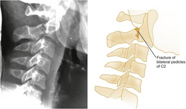
                        Hangman's Fracture 
                        Mechanism: extension
                        Stability: unstable
                        Fracture lines extending through the pedicles of C2 are well visualized
                        Retropharyngeal soft tissue swelling is apparent

                        Tardieu’s Spot
                        在絞扼上方之結膜、黏膜,皮膚造成點狀出血點

                        院內死亡主要原因
                        肺部併發症(吸入性肺炎、肺水腫、急性呼吸窘迫症)

                        2011年1月25日 星期二

                        The Management of Pancreatic Trauma in the Modern Era


                        The Management of Pancreatic Trauma in the Modern Era
                        Surgical Clinics of North America. Volume 87, Issue 6 (December 2007)

                        Diagnosis
                        • Grading system
                        • Serum amylase levels
                        • CT
                        • ERCP
                        • DSS MRCP
                        • Exploratory laparotomy
                        Pancreas Organ Injury Scale of the American Association for the Surgery of Trauma

                        Grade
                        Injury
                        Description
                         I
                        Hematoma
                        Minor contusion without duct injury

                        Laceration
                        Superficial laceration without duct injury
                         II
                        Hematoma
                        Major contusion without duct injury or tissue loss

                        Laceration
                        Major laceration without duct injury or tissue loss
                         III
                        Laceration
                        Distal transection or parenchymal injury with duct injury
                         IV
                        Laceration
                        Proximal transection or parenchymal injury involving ampulla
                         V
                        Laceration
                        Massive disruption of pancreatic head

                        CT findings suspicious for an injury to the pancreas include the following:
                        • hematoma surrounding the pancreas
                        • fluid in the lesser sac
                        • thickening of the left anterior Gerota's fascia.
                        CT scans can also demonstrate parenchymal lacerations or transections of the main pancreatic duct.

                        ERCP is the most reliable method to define continuity of the main pancreatic duct accurately.
                        Grade
                        Description
                        I
                        Normal main pancreatic duct on ERCP
                        IIa
                        Injury to branches of main pancreatic duct on ERCP with contrast extravasation inside the parenchyma
                        IIb
                        Injury to branches of main pancreatic duct on ERCP with contrast extravasation into the retroperitoneal space
                        IIIa
                        Injury to the main pancreatic duct on ERCP at the body or tail of the pancreas
                        IIIb
                        Injury to the main pancreatic duct on ERCP at the head the pancreas

                        Exploratory Laparotomy
                        In those patients who are taken emergently to the operating room for abdominal trauma, pancreatic injuries are diagnosed at exploration and it is important to establish the continuity of the main pancreatic duct.
                        Nonoperative Management
                        If there is no evidence of a ductal injury on fine-cut CT, non-operative management is acceptable, although it may be wise to perform ERCP to establish normal ductal anatomy definitively.
                        Operative Treatment
                        Indications

                        • Peritonitis on physical examination
                        • Hypotension and a positive FAST
                        • Evidence of disruption of the pancreatic duct on fine-cut CT or on ERCP
                        Isolated injuries to the pancreas without ductal involvement

                        General principles and exposure
                        During laparotomy, the initial priorities are control of active hemorrhage and control of gross gastrointestinal contamination.
                         Simple external drainage
                        In the hemodynamically stable patient, pancreatic contusions (AAST grade I), minor capsular injuries, and traumatic pancreatitis can be treated without drainage.
                        Most other injuries require drainage of some sort.
                        Treatment options for isolated pancreatic injuries based on the American Association for the Surgery of Trauma pancreas Organ Injury Scale

                        AAST grade
                        Treatment options
                         I
                        Observation

                        Omental pancreatorrhaphy with simple external drainage
                         II
                        Simple external drainage

                        Omental pancreatorrhaphy and drainage
                         III
                        Distal pancreatectomy ± splenectomy

                        Roux-en-Y distal pancreatojejunostomy
                         IV
                        Pancreatoduodenectomy

                        Roux-en-Y distal pancreatojejunostomy

                        Anterior Roux-en-Y pancreatojejunostomy

                        Endoscopically placed stent

                        Simple drainage in damage control situations
                         V
                        Pancreatoduodenectomy

                        Isolated pancreatic injuries with ductal involvementp1
                        Distal pancreatectomy
                        In the hemodynamically stable patient with an isolated pancreatic injury, especially a child 10 years of age or younger, splenic salvage should be considered.


                        p2
                        If the patient is hemodynamically unstable, an expeditious distal pancreatectomy with splenectomyshould be performed.


                        p3Roux-en-Y distal pancreatojejunostomy is indicated in hemodynamically stable patient who has a transection of the pancreas at the neck or just to the right of the mesenteric vessels and few associated injuries.


                        Combined pancreatoduodenal injuries

                        Pancreatoduodenectomy is indicated when there is extensive trauma to the head of the pancreas, a severe combined pancreatoduodenal injury, or destruction of the ampulla of Vater.
                        • In the hemodynamically stable patient, this procedure can be performed at the time of the original trauma laparotomy.
                        • In most of the patients who are hypothermic, acidotic, or coagulopathic, a damage control procedure is indicated. In this instance, the pancreatoduodenectomy or the reconstruction after a prior pancreatoduodenectomy should be performed at the reoperation.
                        • Control of hemorrhage and gastrointestinal contamination must occur first.
                        p4